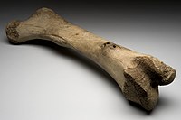
Photo from wikipedia
OBJECTIVE To evaluate the healing of critical size defects (CSDs) in calvaria of rats with osteoporosis induced by ovariectomy and treated with L-PRF associated or not with bovine bone graft… Click to show full abstract
OBJECTIVE To evaluate the healing of critical size defects (CSDs) in calvaria of rats with osteoporosis induced by ovariectomy and treated with L-PRF associated or not with bovine bone graft (XENO). MATERIAL AND METHODS 32 rats underwent a bilateral ovariectomy procedure. After 3 months, one 5-mm in diameter CSD was created in the middle of the calvaria of each animal. In group C, defect was filled with blood clot only. In PRF, XENO and PRF-XENO groups, defects were filled with 0.1 mL of L-PRF, 0.1 mL of XENO and a mixture of 0.1 mL of L-PRF plus 0.1 mL of XENO, respectively. L-PRF compressed clots were used to cover the defects in PRF and PRF-XENO groups. Animals were submitted to euthanasia at 30 postoperative days. Histomorphometric, microtomographic and immunohistochemical analyses were performed. RESULTS PRF-XENO group presented greater amount of neoformed bone (NB) when compared with XENO group, as well as higher expression of Vascular Endothelial Growth Factor (VEGF), Osteocalcin (OCN) and Bone Morphogenetic Protein (BMP-2/4) (p < 0.05). PRF group presented increased amount of NB and higher expression of VEGF, OCN, BMP-2/4 and Runt-related transcription factor 2 (RUNX-2) when compared with group C (p < 0.05). CONCLUSIONS i) the isolated use of L-PRF clot can improve bone neoformation in CSDs in rats with osteoporosis induced by ovariectomy, but seems to lead to decreased amount of bone neoformation when compared to the isolated use of XENO; ii) L-PRF potentiates the healing of XENO in rats with osteoporosis induced by ovariectomy. This article is protected by copyright. All rights reserved.
Journal Title: Clinical oral implants research
Year Published: 2019
Link to full text (if available)
Share on Social Media: Sign Up to like & get
recommendations!