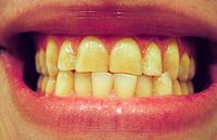
Photo from wikipedia
OBJECTIVES To quantify the neighboring and antagonist teeth migration of a single posterior tooth-missing site within three months using digital scanning and measuring techniques. MATERIAL AND METHODS Intraoral scans (IOS)… Click to show full abstract
OBJECTIVES To quantify the neighboring and antagonist teeth migration of a single posterior tooth-missing site within three months using digital scanning and measuring techniques. MATERIAL AND METHODS Intraoral scans (IOS) were made in 40 patients presenting a single posterior tooth-missing gap and receiving implant therapy. IOS were obtained at the day of and three months after implant surgery rendering a digital baseline model (BM) and a digital follow-up model (FM). Digital models were superimposed using the implant scan body as reference. Antagonist models were processed by the best fit alignment. Dimensional change between anatomical landmarks on neighboring teeth, and that of featuring points on antagonistic teeth were measured using a three-dimensional analysis software. The Mann-Whitney U test was applied to compare the tooth moving distance between the mesial and distal neighboring teeth. The Kruskal-Wallis one-way ANOVA was used to test the difference in dimensional change of tooth-missing site among age subgroups. RESULTS The mean dimensional change of the tooth-missing site was -37.62 ±106.36 μm (median: -28.33 μm, Q25 -72.65/Q75 38.97) mesial-distally and -67.91 ±42.37 μm (median: -61.50 μm, Q25 -88.25/Q75 -36.75) occlusal-gingivally. Eighteen out of 40 mesial neighboring teeth and 24 out of 40 distal neighboring teeth showed migration towards the implants. When patients were grouped according to age, the mesial-distal reduction of the tooth-missing site was significantly larger in patients younger than 30 years compared to those older than 50 years (P<0.05). CONCLUSIONS The dimensions of posterior tooth-missing sites decreased over an observation period of three months.
Journal Title: Clinical oral implants research
Year Published: 2020
Link to full text (if available)
Share on Social Media: Sign Up to like & get
recommendations!