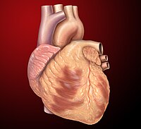
Photo from wikipedia
Papillary muscle (PM) rupture is a rare complication of acute myocardial infarction which carries an excessive mortality rate. Optimal outcomes require rapid diagnosis and prompt surgical referral, and in this… Click to show full abstract
Papillary muscle (PM) rupture is a rare complication of acute myocardial infarction which carries an excessive mortality rate. Optimal outcomes require rapid diagnosis and prompt surgical referral, and in this regard, echocardiography plays a crucial role. Comprehensive echocardiographic examination of the patient with PM rupture consists of identification of the ruptured PM segment, visualization of flail mitral valve segment(s), evaluation of mitral regurgitation severity, and assessment of left ventricular systolic function. This article discusses anatomic and echocardiographic features as well as the surgical management of PM rupture.
Journal Title: Echocardiography
Year Published: 2017
Link to full text (if available)
Share on Social Media: Sign Up to like & get
recommendations!