Photo from wikipedia
Paraneoplastic pemphigus without mucosal lesions in a patient with ovarian cancer Dear Editor, A 48-year-old female with a BRCA2 gene mutation and a family history of breast cancer presented with… Click to show full abstract
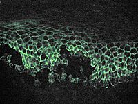
Paraneoplastic pemphigus without mucosal lesions in a patient with ovarian cancer Dear Editor, A 48-year-old female with a BRCA2 gene mutation and a family history of breast cancer presented with a 6-month history of widespread polymorphous cutaneous lesions and severe pruritus. Her lesions consisted of erythematous annular and targetoid papules and plaques on the trunk and extremities; multiple edematous, erythematous wheals on the trunk and forearms; and a focal area of clear-filled vesicles admixed with eroded, erythematous papules on the right dorsal forearm (Fig. 1). She had no erosions, bullae, scale, or crusting of the ocular, oral, or genital mucosa. Prior treatments for her lesions included triple antibiotic ointment, doxycycline, and antihistamines. She also reported taking 20 mg prednisone daily prior to referral to our clinic, at which time she did not have any significant skins lesions except for a few scattered hyperpigmented patches located on her trunk and extremities. A punch biopsy of the thigh revealed eosinophilic spongiosis with intercellular edema in the epidermis, lymphocytic exocytosis, superficial perivascular infiltrate of lymphocytes, and scattered eosinophils. Direct immunofluorescence (DIF) exhibited intercellular IgG and C3, in focal deposits, consistent with pemphigus. A repeat punch biopsy of the abdomen showed similar findings with a vacuolar interface dermatitis associated with eosinophils. DIF for this specimen exhibited granular, pericellular deposition of IgG in the epidermis and discontinuous granular deposition of C3 at the dermal-epidermal junction (Fig. 2). Serum testing showed antibodies to desmoglein-3 (Dsg-3) (170 U/ml). Antibodies targeting desmoglein-1 (Dsg-1) and bullous pemphigoid BP230 were absent. Months after her initial visit, the patient began to experience weight loss and abdominal fullness. Imaging and diagnostic workup revealed metastatic high-grade ovarian serous carcinoma. The constellation of her skin findings along with the discovery of a neoplasm were consistent with paraneoplastic pemphigus. To our knowledge, this is the first report of paraneoplastic pemphigus associated with serous ovarian carcinoma. This is perhaps not surprising given that pemphigus has been associated with both benign and malignant neoplasms. It is primarily seen in relation to lymphoreticular malignancies but has also been observed in some solid tumors. The polymorphic cutaneous lesions observed in this patient reflect the heterogeneous clinical and pathologic presentations of neoplastic-associated pemphigus described in literature including: pemphigus-like, pemphigoid-like, erythema multiforme-like, graft-vs-host disease-like, and lichen planus-like lesions. Importantly, many articles indicate that erosive, progressive, and painful mucositis is uniformly present in patients with paraneoplastic pemphigus, and in its absence, the diagnosis should not be considered. However, our patient with immune-pathologic features consistent with pemphigus did not exhibit clinically evident mucosal lesions. This recognition is of paramount importance given that cutaneous features of paraneoplastic pemphigus may present months before a malignancy is detected and potentially at earlier stages of disease, particularly in tumors such as serous ovarian carcinoma, where the majority of patients are diagnosed with advanced disease. Varied histopathologic findings observed in paraneoplastic pemphigus mirror the heterogenous clinical presentations and
Journal Title: International Journal of Dermatology
Year Published: 2021
Link to full text (if available)
Share on Social Media: Sign Up to like & get
recommendations!