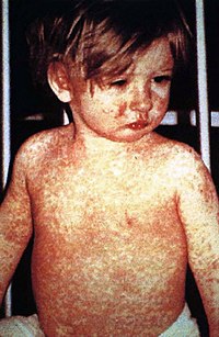
Photo from wikipedia
above the tumor (OR = 11.67, 95% CI 2.40–56.75, P = 0.003) or upper edge of the tumor located in the dermis (OR = 17.78, 95% CI 2.46–128.65, P =… Click to show full abstract
above the tumor (OR = 11.67, 95% CI 2.40–56.75, P = 0.003) or upper edge of the tumor located in the dermis (OR = 17.78, 95% CI 2.46–128.65, P = 0.001) more frequently than non-redcolored ones with adjustment for epidermal change as confounding factor in the Mantel–Haenszel test. It has been reported that the rate of correct diagnosis in pilomatricomas was16%. Especially, pilomatricomas have been occasionally misdiagnosed as epidermoid cyst. Indeed, both pilomatricoma and epidermoid cyst present with red-colored or skin-colored appearance frequently. This study suggests that most pilomatricomas generated through the dermis present red-colored appearance by causing inflammation around themselves. When we encounter red-colored tumors, pilomatricomas should be considered as differential diagnosis as well as epidermoid cyst regardless of skin change overlying the tumor with the understanding of the mechanism to cause red-colored tumors.
Journal Title: International Journal of Dermatology
Year Published: 2022
Link to full text (if available)
Share on Social Media: Sign Up to like & get
recommendations!