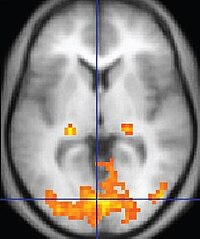
Photo from wikipedia
Magnetic resonance imaging (MRI) has been used to visualize radiofrequency (RF) ablation lesions but the relationship between volumes that enhance in acute MRI and the chronic lesion size is unknown. Click to show full abstract
Magnetic resonance imaging (MRI) has been used to visualize radiofrequency (RF) ablation lesions but the relationship between volumes that enhance in acute MRI and the chronic lesion size is unknown.
Journal Title: Journal of Cardiovascular Electrophysiology
Year Published: 2018
Link to full text (if available)
Share on Social Media: Sign Up to like & get
recommendations!