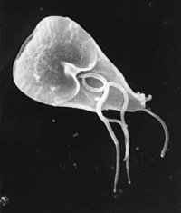
Photo from wikipedia
H.C. Tseng et al., Taiwan, wrote an interesting article about the clinicopathological features of cutaneous protothecosis (JEADV, this issue). For many dermatologists in Europe, ‘protothecosis’ is a completely unknown, very… Click to show full abstract
H.C. Tseng et al., Taiwan, wrote an interesting article about the clinicopathological features of cutaneous protothecosis (JEADV, this issue). For many dermatologists in Europe, ‘protothecosis’ is a completely unknown, very rare skin infection. Even those who are specialists for infectious skin diseases and are very well informed about all kinds of bacterial, viral, fungal and parasitic skin infections must not have seen a single case during several decades of daily routine as a dermatologist. Some standard textbooks of dermatology mention the possibility of cutaneous algae infections in their mycology chapters, but in common, the information you get is very limited and photographs of skin lesions and histopathology are hard to find. Algae are a diverse, polyphyletic group of eukaryote organisms. Not very closely related species from the achlorophyllic genus Prototheca, Chlorella and, very recently from chlorophyllic Desmodesmus sp. were found to cause opportunistic skin infections in humans. Historically even prokaryote cyanobacteria are named ‘blue-green algae’, but they are bacteria. Most algae are found in the oceans, but microalgae are also found in freshwater. Prototheca spp. are a ubiquitous genus of unicellular, achlorophyllous, saprophytic algae. They are frequently found in water (sewage), soil and on plant surfaces. They also can be part of the transient flora of the gastrointestinal tract in animals and humans and can asymptomatically colonize skin and nailbeds. Sporadic infections in dogs, cats and non-domesticated mammals, as well as endemic infections in cows (mastitis), are reported worldwide, but seem to be more frequent in tropical or subtropical countries. According to the literature, human protothecosis was first described in 1964, cited by Hillesheim. Since then, less than 100 cases (including the 20 cases of Tseng’s review in this issue have been reported. Although these algae are ubiquitous saprophytes, most human infections are reported from Asian countries with a tropical or subtropical climate. In humans, accidental inoculation of prototheca (mostly P. wickerhamii) into the skin through wounds or traumatic injury may result in cutaneous infection. Repeated traumatic inoculation is also suspected to be the cause of protothecal bursitis olecrani. Protothecosis is an opportunistic infection found in association with local or systemic immunodeficiency. Growing within the patient’s skin, algae produce inflammatory granulomatous lesions, which have to be differentiated from deep fungal and mycobacterial infections. In cases of severe immunodeficiency, visceral dissemination or even sepsis may follow local cutaneous infection. A suspicion of protothecosis arouses in skin disease, that is clinically and morphologically very likely to be a deep fungal infection with granulomatous, eczematous or ulcerating lesions. In the review by Tseng et al., eczematous lesions of the limbs had been the most frequent clinical sign of protothecosis. The pathogens are detected directly in skin biopsies as typical endosporulating sporangia. Morphologically, they have to be differentiated from fungi like blastomyces, Coccidioides immitis, Paracoccidioides brasiliensis, Cryptococcus neoformans and others (Narayan stain). Prototheca spp. can be cultured on standard media for fungi (without cycloheximide) and blood agar. For special questions, several nucleic acid amplification tests (NAAT) are available. Tseng et al. wrote an interesting paper about the clinical and histopathological features of this rare disease. They analysed 20 cases of pathology confirmed cutaneous protothecosis in Taiwan (the largest case series so far). All patients had some kind of immunodeficiency, lived in rural areas, and most of them had indolent chronic erythematous patches or plaques with pustules and small ulcers typically located on their limbs. A history of trauma in the diseased skin location was positive in one-third of the patients only. Granulomatous skin reactions and sporangia in the dermis were the most important histopathological findings. Unfortunately, because of the retrospective character of this study, identification of prototheca species was successful in two of 20 cases only (P. wickerhamii). Interestingly, there seems to be some unexplained association of protothecosis with scabies. Every 5th patient of Tseng’s study group had scabies too. So, in cases of itchy protothecosis, there should be a suspicion of scabies comorbidity. To learn more about algae as pathogens in human skin disease, it is mandatory to proof infection by histopathology, culture and identification of the infective agent on a species level.
Journal Title: Journal of the European Academy of Dermatology and Venereology
Year Published: 2018
Link to full text (if available)
Share on Social Media: Sign Up to like & get
recommendations!