Photo from wikipedia
Endoscopic treatment of benign bilioenteric anastomosic strictures is challenging because the biliary orifice is not easily accessible after biliary diversion surgery. We report a case of recurrent benign hepaticojejunostomy anastomotic… Click to show full abstract
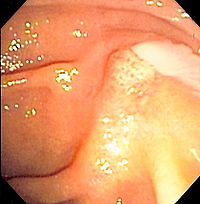
Endoscopic treatment of benign bilioenteric anastomosic strictures is challenging because the biliary orifice is not easily accessible after biliary diversion surgery. We report a case of recurrent benign hepaticojejunostomy anastomotic stricture following pylorus-preserving pancreaticoduodenectomy, treated with percutaneous transhepatic placement, and endoscopic removal of a retrievable, fully covered metal stent with a 50-cm-long lasso. A 41-year-old woman underwent pylorus-preserving pancreaticoduodenectomy for duodenal cancer in 2015 and percutaneous balloon dilation with prolonged catheter interposition for a benign hepaticojejunostomy anastomotic stricture after failed endoscopic treatment in 2016. She developed jaundice secondary to a recurrent anastomotic stricture in 2017. We performed percutaneous transhepatic biliary drainage and obtained cholangiogram (Fig. 1a). The contrast medium did not flow to the jejunum because of a tight hepaticojejunostomy anastomotic stricture. Three days after percutaneous transhepatic drainage, she underwent percutaneous transhepatic placement of a 10 × 3-cm retrievable covered stent with a 50-cm-long distal lasso with five radiopaque markers at 10-cm intervals (M-Intraductal; Standard Sci-Tech Inc, Korea) (Fig. 1b). Before stent insertion, a 0.035-inch guidewire was advanced maximally distally from the anastomotic site for lasso deployment and to obtain enterography images (Fig. 1c). The lasso and stent were sequentially deployed (Fig. 1d,e). Radiographic images were obtained 1 day and 1, 2, 4, 8, and 12 weeks after the procedure to locate the lasso distal tip and check for stent migration. Three months after stent insertion, a conventional gastroscope (GIF-XQ260; Olympus) was advanced to the afferent jejunal limb. The lasso was grasped using rat-tooth forceps, 80 cm from the incisors, and the stent was removed by retracting the forceps within the working channel (Fig. 1f,g). She has not reported recurrent obstructive jaundice for a year.
Journal Title: Journal of Gastroenterology and Hepatology
Year Published: 2019
Link to full text (if available)
Share on Social Media: Sign Up to like & get
recommendations!