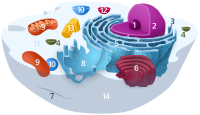
Photo from wikipedia
The actin cytoskeleton is a main component of cells and it is crucially involved in many physiological processes, e.g. cell motility. Changes in the actin organization can be effected by… Click to show full abstract
The actin cytoskeleton is a main component of cells and it is crucially involved in many physiological processes, e.g. cell motility. Changes in the actin organization can be effected by diseases or vice versa. Due to the nonuniform pattern, it is difficult to quantify reasonable features of the actin cytoskeleton for a significantly high cell number. Here, we present an approach capable to fully segment and analyse the actin cytoskeleton of 2D fluorescence microscopic images with a special focus on stress fibres. The extracted feature data include length, width, orientation and intensity distributions of all traced stress fibres. Our approach combines morphological image processing techniques and a trace algorithm in an iterative manner, classifying the segmentation result with respect to the width of the stress fibres and in nonfibre‐like actin. This approach enables us to capture experimentally induced processes like the condensation or the collapse of the actin cytoskeleton. We successfully applied the algorithm to F‐actin images of cells that were treated with the actin polymerization inhibitor latrunculin A. Furthermore, we verified the robustness of our algorithm by a sensitivity analysis of the parameters, and we benchmarked our algorithm against established methods. In summary, we present a new approach to segment actin stress fibres over time to monitor condensation or collapse processes.
Journal Title: Journal of Microscopy
Year Published: 2017
Link to full text (if available)
Share on Social Media: Sign Up to like & get
recommendations!