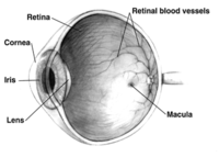
Photo from wikipedia
Retinal pigment epithelium (RPE) is a specialized pigmented monolayer dedicated to retinal support and protection. Given the fact that photoreceptor outer segments are the primary energy resource of RPE metabolism,… Click to show full abstract
Retinal pigment epithelium (RPE) is a specialized pigmented monolayer dedicated to retinal support and protection. Given the fact that photoreceptor outer segments are the primary energy resource of RPE metabolism, it follows that, when photoreceptor function is compromised, RPE cells are impaired and vice versa. In retinitis pigmentosa (RP), genetic mutations lead to a massive degeneration of photoreceptors but only few studies have addressed systematically the consequences of rod and cone death on RPE cells, which, among others, undergo an abnormal organization of tight junctions (TJs) and a compromised barrier function. The biological mechanisms driving these barrier reorganizations are largely unknown. Studies aimed at addressing general and mutation-independent changes of the RPE in RP are relevant to reveal new pathogenic mechanisms of this heterogeneous family of diseases and prospectively develop effective therapeutic strategies. Here, we take advantage of a mouse model of RP in which retinal degeneration is spatially restricted to investigate a possible involvement of inflammatory responses in RPE remodeling. By immunostaining for Zona Occludens-1 (ZO-1), a structural and functional marker of TJs with pleiotropic functions, we found a partial rescue of TJs organization following local restoration of retinal organization, revealing that TJs structure can recover. Since lack of ZO-1 from TJs can alter cell density, we counted RPE cells without finding any differences between degenerated and controls animals, indicating preservation of RPE cells. However, we found an increased number of immune cells adhering to the RPE apical surface and a spatial correlation with areas of abnormal ZO-1 distribution. This suggests that inflammatory processes following photoreceptor degeneration can be responsible for TJs alterations during RP progression and deserve further investigation.
Journal Title: Journal of anatomy
Year Published: 2022
Link to full text (if available)
Share on Social Media: Sign Up to like & get
recommendations!