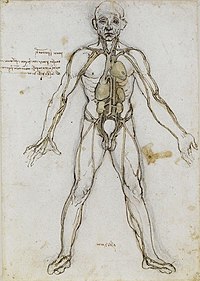
Photo from wikipedia
Understanding how the brain is provided with glucose and oxygen is of particular interest in human evolutionary studies. In addition to the internal carotid arteries, vertebral arteries contribute significantly to… Click to show full abstract
Understanding how the brain is provided with glucose and oxygen is of particular interest in human evolutionary studies. In addition to the internal carotid arteries, vertebral arteries contribute significantly to the cerebral and cerebellar blood flow. The size of the transverse foramina has been suggested to represent a reliable proxy for assessing the size of the vertebral arteries in fossil specimens. To test this assumption, here, we statistically explore spatial relationships between the transverse foramina and the vertebral arteries in extant humans. Contrast computed tomography (CT) scans of the cervical regions of 16 living humans were collected. Cross‐sectional areas of the right and left transverse foramina and the corresponding vertebral arteries were measured on each cervical vertebra from C1 to C6 within the same individuals. The cross‐sectional areas of the foramina and corresponding arteries range between 13.40 and 71.25 mm2 and between 4.53 and 29.40 mm2, respectively. The two variables are significantly correlated except in C1. Using regression analyses, we generate equations that can be subsequently used to estimate the size of the vertebral arteries in fossil specimens. By providing additional evidence of intra‐ and inter‐individual size variation of the arteries and corresponding foramina in extant humans, our study introduces an essential database for a better understanding of the evolutionary story of soft tissues in the fossil record.
Journal Title: Journal of Anatomy
Year Published: 2022
Link to full text (if available)
Share on Social Media: Sign Up to like & get
recommendations!