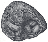
Photo from wikipedia
A 51-year-old female was admitted with acute chest pain. She had experienced intermittent chest pain for more than 2 years. An electrocardiogram showed no acute changes; however, a stress test… Click to show full abstract
A 51-year-old female was admitted with acute chest pain. She had experienced intermittent chest pain for more than 2 years. An electrocardiogram showed no acute changes; however, a stress test revealed ST segment elevation in AVR and diffuse ST segment depression associated with chest pain. A cardiac angiogram showed a tortuous vessel arising form a normal right coronary artery draining into a normal left circumflex (LCx) artery (Fig. 1). Since the origin of the left main coronary artery (LMCA) could not be visualized, a multi-planar computed tomography angiogram was performed which revealed a 50-75% ostial LMCA lesion. The LMCA arose from the left ventricular outflow tract at the level of the left and non-coronary commissures (Figs. 2-4). There were no other lesions in the left anterior descending (LAD) or LCx arteries. The patient underwent an uneventful saphenous vein to LAD bypass. The left internal mammary artery was encased in firm, inflammatory tissue and was foreshortened so that it could not reach the LAD anastomotic site. The patient remains symptom free 1 year following surgery.
Journal Title: Journal of Cardiac Surgery
Year Published: 2017
Link to full text (if available)
Share on Social Media: Sign Up to like & get
recommendations!