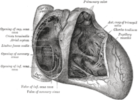
Photo from wikipedia
Lipomatous hypertrophy of the interatrial septum (LHIS), a fatty tumor, is usually diagnosed on both echo and CT/MRI imaging. Cases of LHIS located outside of the interatrial septum are extremely… Click to show full abstract
Lipomatous hypertrophy of the interatrial septum (LHIS), a fatty tumor, is usually diagnosed on both echo and CT/MRI imaging. Cases of LHIS located outside of the interatrial septum are extremely rare and rarer still are these cases large enough to cause symptoms. The clinical literature demonstrates a misunderstanding that fatty tumors outside the intra‐atrial area represent lipomas. However, pathologic understanding of these fatty tumors is clear and is based on microscopic findings.
Journal Title: Journal of Cardiac Surgery
Year Published: 2020
Link to full text (if available)
Share on Social Media: Sign Up to like & get
recommendations!