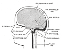
Photo from wikipedia
In an interesting brief report in this issue of the Journal, Feng and his colleagues describe their experience with a valve related to the orifice of the superior caval vein.… Click to show full abstract
In an interesting brief report in this issue of the Journal, Feng and his colleagues describe their experience with a valve related to the orifice of the superior caval vein. They point out that, although remnants of the valves of the embryonic venous sinus are well recognised to persist in postnatal life as the valves of the inferior caval vein and the coronary sinus, it is rare to find such valvar remnants related to the orifice of the superior caval vein. As they correctly explain, the remnants related to the inferior caval vein and the coronary sinus are parts of a complete valvar complex. When first formed, they mark the boundary, during embryonic development, between the systemic venous sinus and the right atrium. In support of this statement, they make reference to a recent study conducted by Hikspoors and colleagues. This work provides a pictorial account of the anatomical changes taking place during embryonic development of the human heart. The beauty of this report, in which one of us (RHA) was privileged to take part, is that the three‐dimensional datasets used by the investigators have now been made available as interactive portable document format files. These files can be interrogated and reconstructed by the interested reader. By selecting the appropriate segmented components of the developing heart, it is possible to appreciate how, at the early stages of development, the systemic venous tributaries are bilaterally symmetrical, with tributaries from the umbilical, vitelline, and cardinal systems draining into the right and left sinus horns (Figure 1A). At this initial stage, furthermore, each of the sinus horns is in direct continuity with the atrial component of the developing primary heart tube. This process of development is described as stage 12 within the system initially proposed by the Carnegie Institute, where the serial sectioned human embryos from which the reconstructions were made are still archived. These processes take place during the fifth week of development. By the next stage of development, or Carnegie stage 13, a remarkable change has occurred. The systemic venous tributaries have remodelled in such a way that the left sinus horn has become incorporated within the left atrioventricular junction. As a result, the overall systemic venous sinus then drains exclusively to the right side of the developing atrial chambers (Figure 1B). It is this rightward shift of the systemic venous tributaries that sets the scene for subsequent atrial septation. The remodelling uses as a fulcrum the so‐called dorsal mesocardium. This is the stalk, which anchors the developing heart to the pharyngeal mesenchyme. It is the connection provided through the dorsal mesocardium that is used by the pulmonary vein as it canalises within the pharyngeal mesenchyme, gaining its entry to the developing left atrium. It is only subsequent to the rightward shift of the systemic venous sinus that it becomes possible to recognise the valves that guard its entrance to the remainder of the developing morphologically right atrium. After this initial transfer, the left sinus horn remains a significant channel, with the venous valves themselves forming an obvious landmark when viewed from the cavity of the right atrium (Figure 2). This arrangement continues to term in animal species such as the mouse, in which the left sinus horn persists as the left superior caval vein (Figure 3). In normal human development, however, the left sinus horn diminishes markedly in size. It persists only as the coronary sinus, which then serves as the conduit for drainage of the larger part of the venous return from the heart itself. In keeping with the diminution in size of the left sinus horn, in the human heart there is comparable diminution in
Journal Title: Journal of Cardiac Surgery
Year Published: 2022
Link to full text (if available)
Share on Social Media: Sign Up to like & get
recommendations!