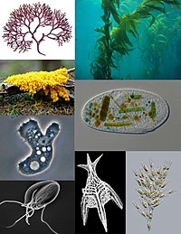
Photo from wikipedia
Decades ago, the major tools that evolutionary phycologists employed to infer relationships among eukaryotic algae included comparative ultrastructure of cell division processes and the flagellar apparatus of motile cells. Many… Click to show full abstract
Decades ago, the major tools that evolutionary phycologists employed to infer relationships among eukaryotic algae included comparative ultrastructure of cell division processes and the flagellar apparatus of motile cells. Many of the phylogenetic conclusions arising from comparative ultrastructure have since been confirmed by modern analyses that employ genetic sequences. For example, similarities in ultrastructural features of mitosis and cytokinesis in particular green algae (Fig. 1) to those of land plants revealed relationships that were later substantiated by molecular data. These observations primarily involved comparative assessment of the positions of microtubule assemblages, i.e., distinguishing cytokinetic microtubules arrayed parallel to a developing cell plate (phycoplast) from microtubules positioned perpendicular to a developing cross wall (phragmoplast). In recent work, technological advancements in the use of fluorescence microscopy, and particularly immunofluorescence microscopy, have allowed relatively rapid assessment of evolutionarily significant aspects of cell division, thereby facilitating comparative studies. For example, a comparative immunofluorescence approach allowed Doty et al. (2014) to propose the emergence of plant cytokinesis involving a phragmoplast prior to the diversification of the more structurally complex lineages of the charophycean green algae. In this and other cases, the more time-consuming methods previously used to fix, embed, and section algal materials for inspection by transmission electron microscopy (e.g., Fig. 1) have largely been superseded by such newer approaches. In contrast, assessing comparative structural features of the eukaryotic flagellar apparatus, which remains highly useful in evolutionary studies, yet involves diverse ultrastructural features in addition to microtubules, still requires the use of transmission electron microscopy. The Heiss et al. article “The flagellar apparatus of the glaucophyte Cyanophora cuspidata,” published in this issue, demonstrates why ultrastructure is still needed and at the same time provides an outstanding example of an ultrastructural analysis that has employed state-ofthe-art methods to approach long-standing issues. The unicellular flagellate Cyanophora (Fig. 2) represents the enigmatic algal lineage informally known as the glaucophytes, which, though not diverse, are significant for being related in some way to the modern, diverse lineages informally known as the red algae and the green algae. Diversification of these three algal lineages is central to ongoing controversies surrounding the single versus multiple origin(s) of primary plastids. Because they possess blue-green plastids (see Fig. 2) that retain a carboxysome-like structure and a surface peptidoglycan layer visible with the use of TEM–presumably derived from the cell wall of a cyanobacterial ancestor–glaucophytes suggest early stages in the evolution of primary plastids. Consequently, glaucophytes have attracted the attention of phycologists who have employed cellular, biochemical, and molecular approaches to better understand their relationships. Kugrens et al. (1999), for example, published an important ultrastructural analysis of Cyanophora biloba (in the Journal of Phycology), Abdel-Ghany et al. (2000) explored evolutionary relationships of C. paradoxa kinesin-like protein components (also in J. Phycol.), and Price et al. (2012) reported a phylogenomic analysis of transcriptomic and draft genomic data from C. paradoxa. Heiss et al. do not employ their new ultrastructural data to take a particular evolutionary position, but these authors have produced a technologically proficient analysis and a comprehensive compilation of the relevant systematic literature, both of which will enable future progress. One of the most impressive aspects of the Heiss et al. study is its technical excellence. Since the transmission electron microscope first became available in the 1930s, and Hall and Claus published a first TEM study of Cyanophora in 1963, many highquality ultrastructural studies of algae have been published. Some examples of technically exceptional TEM of unicellular algal flagellates published in the Journal of Phycology during this time are the Lembi (1975) study of the Carteria flagellar apparatus, and the Pearson and Norris (1975) study of Pyramimonas, which included spectacular images of J. Phycol. 53, 1117–1119 (2017) © 2017 Phycological Society of America DOI: 10.1111/jpy.12585
Journal Title: Journal of Phycology
Year Published: 2017
Link to full text (if available)
Share on Social Media: Sign Up to like & get
recommendations!