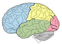
Photo from wikipedia
Reported brain abnormalities in anatomy and function in patients with narcolepsy with cataplexy led to a project based on qualitative electroencephalography examination and analysis in an attempt to find a… Click to show full abstract
Reported brain abnormalities in anatomy and function in patients with narcolepsy with cataplexy led to a project based on qualitative electroencephalography examination and analysis in an attempt to find a narcolepsy with cataplexy‐specific brain‐derived pattern, or a sequence of brain locations involved in processing humorous stimuli. Laughter is the trigger of cataplexy in these patients, and the difference between patients and healthy controls during the laughter should therefore be notable. Twenty‐six adult patients (14 male, 12 female) suffering from narcolepsy with cataplexy and 10 healthy controls (five male, five female) were examined. The experiment was performed using a 256‐channel electroencephalogram and then processed using specialized software built according to the scientific research team's specifications. The software utilizes electroencephalographic data recorded during elevated emotional states in participants to calculate the sequence of brain areas involved in emotion processing using non‐linear and linear algorithms. Results show significant differences in activation (pre‐laughter) patterns between the patients with narcolepsy and healthy controls, as well as significant similarities within the patients and the controls. Specifically, gyrus orbitalis, rectus and occipitalis inferior are active in healthy controls, while gyrus paracentralis, cingularis and cuneus are activated solely in the patients in response to humorous audio stimulus. There are qualitative electroencephalographic‐based patterns clearly discriminating between patients with narcolepsy and healthy controls during laughter processing.
Journal Title: Journal of Sleep Research
Year Published: 2017
Link to full text (if available)
Share on Social Media: Sign Up to like & get
recommendations!