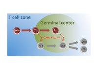
Photo from wikipedia
Background Rheumatoid arthritis (RA) is a highly disabling autoimmune disease. T lymphocyte subsets imbalance causeing immune dysfunction is essential stages in the occurrence and development of RA diseases. Recent studies… Click to show full abstract
Background Rheumatoid arthritis (RA) is a highly disabling autoimmune disease. T lymphocyte subsets imbalance causeing immune dysfunction is essential stages in the occurrence and development of RA diseases. Recent studies show that the interaction of both follicular helper T (Tfh) cells and follicular regulatory T (Tfr) cells are important frontier scientific direction to maintain autoimmune tolerance, and Tfr/Tfh balance may play a pivotal role in the formation of lymphoid germinal center and the production of autoantibodies[1-2]. Objectives The aim of this study was to explore the clinical characteristic of peripheral follicular T cell subsets in patients with RA and healthy individuals,and the effects of Tfr/Tfh balance on autoantibody formation and disease activity. Searching for new immunomodulatory targets from it. Methods The study included 26 patients with a diagnosis of RA according to the 1987 revised criteria of the American College of Rheumatology and 17 healthy individuals as control group. All follicular T cell subsets from them were assessed by flow cytometry(Figure1). Using isotypic controls to distinguish follicular T cell subsets that were not clearly clustered. In addition, we measured all peripheral lymphocyte subsets in RA patients by Flow Cytometry. IgG, IgA, IgM,RF were measured using Turbidimetric inhibition immuno assay. Anti-CCP was measured using ELISA. We also collected relevant clinical information and made DAS28 score. Results (1)Compared with healthy controls, the proportions of CD4+CXCR5+PD-1highTfh cells were higher in RA patients (P=0.029), In contrast, patients with RA had much lower level of CD25+CXCR5+FoxP3+Tfr cells (P=0.010). And there were significant differences of Tfr/Tfh between these two groups (P=0.000). (2)Among 26 RA patients, there was obvious correlation between Tfr/Tfh and the DAS28 value(r=0.422, P=0.032). But there was no correlation between Tfr/Tfh and ESR(P>0.05). Correlation between CD4+CD25+ FoxP3+Treg cells and Tfr cells was also analyzed(r=0.722, P=0.000)(Figure2). (3)We have not found correlation between Tfr/Tfh and autoantibodies maybe for the small sample content. Conclusion Treg cells have a negative immunomodulatory effect on inflammatory response, the high correlation between Tfr cells and Treg cells indicates that Tfr cells may come from directional transformation of Treg. There is a Tfr/Tfh imbalance in RA patients, which suggests a potential mechanism of RA disease severity. However, more samples are needed to confirm whether it is related to the production of autoantibodies to affect disease activity. References [1] Wing J B, Tekgüc Murat, Shimon S. Control of germinal center responses by T-follicular regulatory cells[J]. Frontiers in Immunology, 2018, 9:1910-1922. [2] Moschovakis G L, Bubke A, Friedrichsen M, et al. T cell specific CXCR5 deficiency prevents rheumatoid arthritis[J]. Scientific Reports, 2017, 7(1):8933-8946.Figure 1 Follicular T lymphocytes from RA patients and healthy controls were analyzed through staining for CD3, CD4, CXCR5, CD45RA and PD-1 as Tfh cells, andCD3, CD4, CD25, CXCR5,CD45RAandFoxP3 as Tfr cells. Using isotypic control IgG1 to distinguish follicular T cell subsets that were not clearly clustered. Numbers indicate the percentage of cells of last gate.Figure 2 The percentage of circulating follicular T lymphocyte subsets in patients with RA and healthy controls (*p<0.05, ***p<0.001). Correlation between Treg cells and Tfr cells, and correlation between Tfr/Tfh and the DAS28. Disclosure of Interests None declared
Journal Title: Annals of the Rheumatic Diseases
Year Published: 2019
Link to full text (if available)
Share on Social Media: Sign Up to like & get
recommendations!