Photo from wikipedia
Summary An 18 month old boy presented with right monoplegia and intermittent ataxia. This occurred five months post primary varicella infection. His CT brain revealed low dense lesion in the… Click to show full abstract
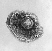
Summary An 18 month old boy presented with right monoplegia and intermittent ataxia. This occurred five months post primary varicella infection. His CT brain revealed low dense lesion in the basal ganglia, a region associated with vasculopathy post varicella infection. The child showed motor recovery with physiotherapy, antiviral, steroid and aspirin. He developed chorea, a manifestation of basal ganglion dysfunction. Case description An 18 month male presented to the emergency department, with inability to use his right hand since the morning and they noticed that he had difficulty crawling without history of trauma or fever. He had primary varicella infection five months earlier, and parents noticed intermittent unsteady gait for the preceding couple of weeks. He had measles mumps and rubella vaccination four months prior to his presentation and had hand foot and mouth disease a month later. He had a simple febrile convulsion four weeks beforehand and was treated as an outpatient in accordance with local guidelines. He attained his normal milestones with no delay, he crawled at one year and walked at 14 months, and he has 20 words with understanding of 3 steps command and respond appropriately. The delivery was full term vaginal delivery with no complications. On initial clinical examination he had no facial asymmetry or any other cranial nerve palsies. There was slight movement against gravity in the right upper limb compared to the left one and his right hand was in continuous fist. Tone in the right upper limb increased compared to the left upper limb with normal reflexes. His gait was normal with no tendency to fall to either side. Extensive laboratory investigations were done, full blood count initially showed leucocytosis with high eosinophilia. CT scan was done and it showed subtle low attenuation in the left globus pallidus. In addition, MRI under general anaesthesia demonstrated a small area of diffusion restriction in the left globs pallidus which indicated ischemia. MR angiography revealed narrowing of anterior cerebral artery and middle cerebral artery compared to the right side. Lumbar puncture was done and it was negative for varicella zoster PCR. Discussion Cerebral complications post varicella infection is a cause of stroke in children. Detailed history of previous infection is essential in making a clinical diagnosis. One literature review states that 44% of cerebrovascular event in children is a consequence/associated with varicella cerebral complications.
Journal Title: Archives of Disease in Childhood
Year Published: 2019
Link to full text (if available)
Share on Social Media: Sign Up to like & get
recommendations!