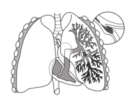
Photo from wikipedia
Pulmonary embolism (PE) is frequently encountered in the emergency department. Syncope, often as a consequence of impending haemodynamic collapse, is associated with increased mortality. While loss of consciousness owing to… Click to show full abstract
Pulmonary embolism (PE) is frequently encountered in the emergency department. Syncope, often as a consequence of impending haemodynamic collapse, is associated with increased mortality. While loss of consciousness owing to cerebral hypoperfusion and reduced left ventricular preload is a common cause of collapse with large volume PE, other syndromes can also cause neurological deficit in thromboembolic disease. Here, we describe a case of a woman in her 60s, presenting to the emergency department with features of high-risk PE. During clinical examination, the patient collapsed and became unresponsive with a Glasgow Coma Scale of 4/15 despite normal haemodynamics. Neurological signs were noted and CT revealed evidence of a large territory cerebral infarction. Further cardiovascular investigations identified a grade 4 patent foramen ovale. We describe a challenging case of established venous thromboembolism complicated by paradoxical embolism, highlighting the importance of thorough clinical examination and investigation and discuss the current evidence base of treatments.
Journal Title: BMJ Case Reports
Year Published: 2022
Link to full text (if available)
Share on Social Media: Sign Up to like & get
recommendations!