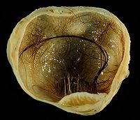
Photo from wikipedia
© BMJ Publishing Group Limited 2022. No commercial reuse. See rights and permissions. Published by BMJ. DESCRIPTION A 35week gestational age woman presented with right lateral neck swelling, previously detected… Click to show full abstract
© BMJ Publishing Group Limited 2022. No commercial reuse. See rights and permissions. Published by BMJ. DESCRIPTION A 35week gestational age woman presented with right lateral neck swelling, previously detected in prenatal ultrasound at 24 weeks of gestation. Elective endotracheal intubation was performed in the first minute of life via ex utero intrapartum (EXIT) procedure. During the first day of life, already in the neonatal intensive care unit, the neonate developed severe respiratory distress, with haemodynamic instability, requiring an increase in the FiO2 up to 100% (previously FiO2 was 30%), and a single dose of surfactant (200 mg/kg) was administrated. On laboratory tests, neoplastic markers, such as alphafetoprotein and human betachoriogonadotropin, were within the normal range. Preoperative ultrasound scan and MRI were performed at the second and third days of life, respectively. MRI revealed an exuberant expansive cervical lesion with welldefined limits centred on the visceral space of the neck with bilateral extension, with slight laterality to the right, resulting in a marked mass effect on the airway and the main cervical vascularnervous axis. The tumour had intermediate T1 signal and T2 moderate–high signal, with multiple lobulations and intrinsic septations, and also intense and relatively homogeneous contrast enhancement (figure 1). After a careful multidisciplinary discussion between paediatric surgery, anaesthesiology, endocrinology, neonatology and radiology, the mass was excised on the fifth day of life, through a transverse cervical incision. To plan the ideal surgical approach, imaging evaluation of the extension of the lesion and its relationship with adjacent structures, such as the trachea, oesophagus and neck vessels, was crucial. A safe airway, that was already established since birth, and haemodynamic monitoring during the procedure were the main factors to consider regarding anaesthetic management. The intraoperative finding consisted of a heterogeneous capsulated cervical tumour measuring 7×5 cm, accompanied by an absent thyroid gland (figure 2). Histopathologic examination showed that the tumour was composed of a variety of tissues from all three germ cell layers, with thyroid tissue also present. There were no signs of immature components or signs of malignancy, consistent with the diagnosis of mature thyroid teratoma. Tight analytical control of thyroid function was performed since the surgery, and on the third postoperative day, levothyroxine (25 μg/day oral) was started due to subclinical hypothyroidism. On the eighth postoperative day, the dose of levothyroxine was adjusted to 12.5 μg/day, which is the current dose. The neonate had the endotracheal tube until the 12th day of life when he was electively extubated to CPAP, and since the 17th day of life has remained without supplemental oxygen therapy. She was discharged from the hospital after 23 days, with favourable clinical evolution. The followup ultrasound, 6 months later, showed no evidence of recurrence. Cervical lesions presenting in the fetal period are often large and may have various aetiologies, with cervical lymphangiomas and cervical teratomas being the two most common. Teratomas are germ cell tumours that contain derivatives of all three germ layers and may originate anywhere along midline and paramidline in both gonadal and extragonadal sites. The head and Figure 1 MRI, (A) axial T2weighted image, (B) axial T1weighted image, (C) axial T1weighted image fat sat G+, (D) coronal T2weighted image, (E) coronal T1weighted image G+ and (F) coronal T1weighted image fat sat G+ showing exuberant expansive cervical lesion with welldefined limits centred on the visceral space of the neck with bilateral extension, with slight laterality to the right, extending superiorly from the plane of the right zygomatic arch to the plane of the sternal notch inferiorly, with a slight insinuation in the superior mediastinum; shows T1 intermediate signal and T2 moderatehigh signal, with multiple lobulations and intrinsic septations, and also intense and relatively homogeneous contrast enhancement, with a preponderant solid lesion and abundant vascular pathways within it; results in marked mass effect, in particular on the airway and the main cervical vascularnervous axis.
Journal Title: BMJ Case Reports
Year Published: 2022
Link to full text (if available)
Share on Social Media: Sign Up to like & get
recommendations!