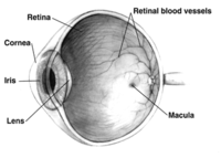
Photo from wikipedia
Background To compare the long-term clinical evolution of highly myopic eyes with vertical oval-shaped dome associated with or without untreated serous retinal detachment. Methods Twenty-eight patients with high myopia (40… Click to show full abstract
Background To compare the long-term clinical evolution of highly myopic eyes with vertical oval-shaped dome associated with or without untreated serous retinal detachment. Methods Twenty-eight patients with high myopia (40 eyes) with smooth macular elevations related to a vertical oval-shaped dome were recruited. Serous retinal detachment was investigated; 11 eyes had persistent submacular fluid (study group) and 29 eyes lacked submacular fluid (control group). All patients underwent complete ophthalmological examinations, including optical coherence tomography at baseline every 6 months for 2 years. Fluorescein angiographies were performed in cases with serous retinal detachment to rule out choroidal neovascularisation. Results There were no statistical differences in baseline age, sex, spherical equivalence or axial length between the two groups. Serous retinal detachment always occurred at the top of the inward macular incurvation. Moreover, no statistically significant differences in mean best-corrected visual acuity were observed during the 24-month follow-up period in the study and control groups and between the two groups at all time points. The mean central foveal thickness was significantly higher in the study group at each visit (p=0.001, in all cases). At the final follow-up visit, complete resolution of the serous retinal detachment was achieved in 1 of the 11 study group’s eyes. Conclusions Serous retinal detachment is a complication associated with vertical oval-shaped domes that seems to remain stable in terms of visual function over time without treatment.
Journal Title: British Journal of Ophthalmology
Year Published: 2018
Link to full text (if available)
Share on Social Media: Sign Up to like & get
recommendations!