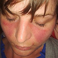
Photo from wikipedia
Background The autoimmune disease systemic lupus erythematosus (SLE) occurs more frequently in patients of African descent with high morbidity and mortality. Current SLE diagnostic criteria including antinuclear antibody (ANA) reactivity… Click to show full abstract
Background The autoimmune disease systemic lupus erythematosus (SLE) occurs more frequently in patients of African descent with high morbidity and mortality. Current SLE diagnostic criteria including antinuclear antibody (ANA) reactivity are derived largely from non-African populations. This study characterises ANA reactivity patterns and relates them to SLE clinical presentation in Black African patients. Methods Sera from Black participants (61 patients with SLE and 100 controls) aged 1–81 years were analysed for reactivity against the antigens: uridine 1-ribonuclear protein, Smith uridine-1-5 ribonuclear protein antigen, soluble substance-A, recombinant Ro-52, soluble substance-B, Scl-70, cytoplasmic histidyl-tRNA synthetase antigen, proliferating cell nuclear antigen (PCNA), nucleosomes, ribonuclear P-protein, antimitochondrial antibody M2 (AMA-M2), histones, double-stranded DNA (dsDNA), centromere protein B and polymyositis–sclerosis overlap antigen. Findings A significantly higher proportion (97%) of the 61 patients with SLE had detectable autoantibody reactivity compared with 15% of the 100 controls (p<0.001). The highest frequencies of autoantibody reactivity in patients with SLE were against the dsDNA antigen (41%) and PCNA (54%). Anti-PCNA and anti-dsDNA reactivity were mutually exclusive (p<0.001) giving rise to two distinct groups of Black African patients with SLE. The first group (n=25) had reactivity profiles consistent with international standard SLE definitions, including anti-dsDNA reactivity, and was 13 times more likely to present with joint symptoms. The larger, second group (n=34), characterised by anti-PCNA and anti-AMA-M2 reactivity, was nine times more likely to present with only cutaneous symptoms. Interpretation Our study demonstrates a need to extend autoantibody panels to include anti-PCNA in the diagnostic process of Black African patients and further refine the predictive values of the reactivity to different antigens to differentiate SLE syndromes in African populations.
Journal Title: BMJ Global Health
Year Published: 2018
Link to full text (if available)
Share on Social Media: Sign Up to like & get
recommendations!