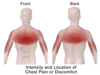
Photo from wikipedia
A 73-year-old woman presented to the ED with non-radiating right sided chest pain, since 1 week. The pain was progressively worsening and was not associated with vomiting and there was no… Click to show full abstract
A 73-year-old woman presented to the ED with non-radiating right sided chest pain, since 1 week. The pain was progressively worsening and was not associated with vomiting and there was no preceding history of trauma. She denied prior episode of chest pain. History was significant for hypertension, hyperlipidaemia and iron deficiency anaemia. There was no history of coughing or smoking. Physical examination and ECG were unremarkable. A frontal chest radiograph was obtained (figure 1). Figure 1 Frontal chest radiograph. Which organ is the most …
Journal Title: Emergency Medicine Journal
Year Published: 2017
Link to full text (if available)
Share on Social Media: Sign Up to like & get
recommendations!