Photo from wikipedia
© Author(s) (or their employer(s)) 2023. No commercial reuse. See rights and permissions. Published by BMJ. INTRODUCTION The healthcare burden posed by aortic stenosis (AS) requires no elaboration. Although it… Click to show full abstract
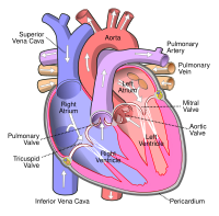
© Author(s) (or their employer(s)) 2023. No commercial reuse. See rights and permissions. Published by BMJ. INTRODUCTION The healthcare burden posed by aortic stenosis (AS) requires no elaboration. Although it is a familiar disease entity that is delineated by a relatively straightforward clinical paradigm, there are still unresolved questions that can make optimal diagnosis, management and followup challenging in some patients. In recent years, computed tomography (CT) has become an indispensable tool in cardiology in view of its excellent spatial resolution, low doses of ionising radiation and widespread availability. Its application in AS is no exception. Cardiac CT may aid in the assessment of native aortic valve (AV) disease, facilitate preprocedural planning for valve interventions and improve the assessment of prosthetic valve dysfunction and structural valve degeneration. This Education in Heart piece will take you through the above, considering at each stage the advantages and disadvantages of CT and the relevant evidence base.
Journal Title: Heart
Year Published: 2023
Link to full text (if available)
Share on Social Media: Sign Up to like & get
recommendations!