Photo from wikipedia
Introduction/Background Ovarian vein thrombosis (OVT) is a rare medical condition most often seen in the immediate postpartum period. OVT has been reported in approximately 0.05–0.18% of vaginal births and in… Click to show full abstract
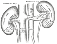
Introduction/Background Ovarian vein thrombosis (OVT) is a rare medical condition most often seen in the immediate postpartum period. OVT has been reported in approximately 0.05–0.18% of vaginal births and in 2% of births by Caesarean section. Anticoagulation is the main therapy used to alleviate symptoms Methodology A 23-year-old female (gravida 0, para 0) presented to obstetrics and gynecology department with symptoms of amenorrhea for 15 week 6 days and intermittent abdominal pain. One month before admission, she was tested positive for urine HCG. Results Laboratory data revealed a b-HCG was elevated (over 200,000 mIU/ml). A transvaginal ultrasound scan revealed a bulky uterus with honeycomb pattern in the endometrial cavity (figure 1). She subsequently underwent a dilatation and curettage. Following this, b-HCG level fell to 43,996.2 mIU/ml. 3 months later, Her b-HCG level again started rising, reaching a level of 138,103.3 mIU/ml. A transvaginal ultrasound scan revealed the right adnexa showed a 9.1 × 5.9 cm multilobular and mixed echogenicity (figure 2a-b). A CT with intravenous contrast revealed multilobulating & tortuous tubular hypodense mass with strong enhencing septae & peripheral wall at right adnexa and extent of the right ovarian vein thrombus which did not reveal other abnormlity (figure 2c). she was started on methotrexate chemotherapy combined with oral anticoagulation with rivaroxaban immediately. 2 weeks later, the b-HCG level showed a fall to 12,364.1 mIU/ml. After several cycles of chemotherapy, the b-HCG level reached a normal level of 2.6 mIU/ml. Conclusion OVT is a rare disorder commonly associated to the peripartum period. But, there was no report of OVT in GTN patients. OVT with only acute abdominal pain is unlikely to be suspected in GTN patients, and thus is found incidentally on CT for metastatic evaluation. Therefore, it is important to have a CT scan if GTN patients have symptoms such as abdominal pain or dyspnea. Disclosure Nothing to disclose Abstract EP1138 Figure 1 Transvaginal ultrasound a-b) uterine, c) Right anexa, d) Left adnexa Abstract EP1138 Figure 2 Transvaginal ultrasound a) Endometrium. b) Right adnexa, c) CT image (OVT)
Journal Title: International Journal of Gynecological Cancer
Year Published: 2019
Link to full text (if available)
Share on Social Media: Sign Up to like & get
recommendations!