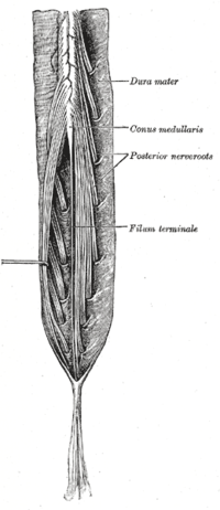
Photo from wikipedia
Although it has long been stated that the level of spinal cord termination varies depending on the size of the dog, the evidence for this remains limited. The aim of… Click to show full abstract
Although it has long been stated that the level of spinal cord termination varies depending on the size of the dog, the evidence for this remains limited. The aim of this study is to investigate the position of the conus medullaris (CM) and dural sac (DS) in a population of dogs of varying size. MRIs of the thoracolumbosacral spine of 101 dogs were included. The location of CM and DS was determined on sagittal T2-weighted images and T1-weighted images, respectively, by three independent observers. The bodyweight and the back length were used as markers of size. Regression analysis showed that the termination point of the CM had a statistically significant relationship with bodyweight (R2=0.23, P<0.05). Although not statistically significant (P=0.058), a similar relationship was found between CM and back length (R2=0.21). No statistically significant relationship was found between the termination point of the DS and bodyweight (P=0.24) or back length (P=0.19). The study confirms the terminal position of the CM is dependent on size, with a more cranial position with increasing size; however, the termination point of DS remains constant irrespective of dog size.
Journal Title: Veterinary Record
Year Published: 2019
Link to full text (if available)
Share on Social Media: Sign Up to like & get
recommendations!