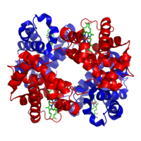
Photo from wikipedia
In the present review, examples are provided illustrating the application of resonance Raman microscopy to heme protein single crystals to highlight the artifacts induced by the crystallization process or the… Click to show full abstract
In the present review, examples are provided illustrating the application of resonance Raman microscopy to heme protein single crystals to highlight the artifacts induced by the crystallization process or the conformational alteration induced by cooling. Moreover, the structural information determined from the RR spectra of heme proteins in solution and crystals is compared to that obtained from their X-ray structures to show how the combined spectroscopic/crystallographic approach is a powerful weapon in the structural biologist’s armamentarium.
Journal Title: Journal of Porphyrins and Phthalocyanines
Year Published: 2019
Link to full text (if available)
Share on Social Media: Sign Up to like & get
recommendations!