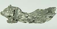
Photo from wikipedia
Background The use of gadolinium-based contrast agents (GBCAs) is linked to gadolinium retention in the skeleton of healthy individuals. The mechanism of gadolinium incorporation into bone tissue is not fully… Click to show full abstract
Background The use of gadolinium-based contrast agents (GBCAs) is linked to gadolinium retention in the skeleton of healthy individuals. The mechanism of gadolinium incorporation into bone tissue is not fully understood and requires spatially resolved analysis to locate the gadolinium. Purpose To compare the quantitative distribution of gadolinium retained over time in rodent femur following the administration of gadodiamide and gadobutrol at three different time points. Materials and Methods In this animal study conducted between May 2018 and April 2020, 108 9-week-old healthy rats were repeatedly injected with either gadodiamide, gadobutrol, or saline solution and were killed 1, 3, or 12 months after the last injection. The femurs of six female and six male rats per each group and time point were collected. Quantitative elemental imaging of gadolinium in longitudinal thin sections was performed on one sample per sex with use of laser ablation inductively coupled plasma mass spectrometry (ICP-MS). Gadolinium concentration was determined with use of ICP-MS on the samples of all animals (six per group). Mann-Whitney U tests were applied on pairwise comparisons to determine potential sex effect and GBCA effect on gadolinium concentrations. Results The highest gadolinium retention was observed in the gadodiamide group (concentration, 97-200 nmol · g-1), exceeding the mean concentration in the gadobutrol group (6.5-17 nmol · g-1). However, the gadolinium distribution pattern was similar for both contrast agents, showing prominent gadolinium retention at endosteal surfaces, in the bone marrow, and in small tissue pores. Gadolinium distribution in cortical bone changed over time, initially showing a thin rim of higher concentration close to the periosteum, which appeared to grow wider and move toward the interior of the femur over 1 year. Conclusion For both gadolinium-based contrast agents, gadolinium retention in rat bone was initially located close to the periosteum and bone cavities and changed with bone remodeling processes. The relevance to long-term storage of gadolinium in humans remains to be determined. © RSNA, 2022 Online supplemental material is available for this article.
Journal Title: Radiology
Year Published: 2022
Link to full text (if available)
Share on Social Media: Sign Up to like & get
recommendations!