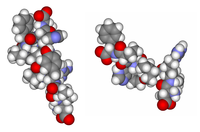
Photo from wikipedia
Vitamin D3 has been proposed as a potential treatment to improve muscle atrophy in sarcopenic patients. Several lines of evidence suggested a possible crosslink between vitamin D and renin-angiotensin system… Click to show full abstract
Vitamin D3 has been proposed as a potential treatment to improve muscle atrophy in sarcopenic patients. Several lines of evidence suggested a possible crosslink between vitamin D and renin-angiotensin system contributing to skeletal muscle atrophy in response to chronic diseases in aging population. Nevertheless, effects of vitamin D and its metabolite on skeletal muscle cells challenged with angiotensin II remain unclear. Therefore, the effects of cholecalciferol (VD3) and calcitriol (1,25VD3) on the ubiquitin-proteasome pathway in angiotensin II-induced skeletal muscle cell atrophy were investigated. Differentiated C2C12 skeletal muscle cells (myotubes) were used as in vitro study model. Myotubes were assigned into vehicle (veh), 1 μM angiotensin II (AII), 1 μM angiotensin II+100 nM cholecalciferol (AII+VD3), 1 μM angiotensin II+1 nM calcitriol (AII+1,25VD3) and 1 μM angiotensin II+10 μM losartan (AII+Lo) groups. Myotubes were applied with angiotensin II on day 4-6, while VD3 and 1,25VD3 were treated on day 5-6 of the differentiation phase. For AII+Lo group, losartan was administered at 2 h before angiotensin II at day 4 of the differentiation period. Cells were harvested at day 7 of differentiation phase for morphological and biochemical analyses. Angiotensin II resulted in significantly decreased myotube diameter and tended to increase protein expression of the transcription factor; FoxO3a and ubiquitin ligases; MURF-1 and atrogin-1, the major muscle protein catabolism pathway when compared with veh-treated group. Treatment of VD3 and losartan could protect angiotensin II-induced myotube atrophy. In contrast, 1,25VD3 accelerated muscle protein breakdown in myotube-challenged with angiotensin II. Protein expression of FoxO3a, MuRF1 and atrogin-1 was significantly up-regulated in AII+1,25VD3 group compared to veh, AII+VD3 and AII+Lo-treated groups. By which these occurs were accompanied with the overexpression of vitamin D receptor in myotube co-treated with angiotensin II and 1,25VD3. Herein, differential effects of VD3 and 1,25VD3 in angiotensin II-induced myotube atrophy were demonstrated. Administration of VD3 resulted in attenuated degree of muscle loss-induced by angiotensin II, while 1,25VD3 possessed the negative impact to skeletal muscle protein metabolism. Collectively, vitamin D3 supplement may be considered as an alternative adjuvant to alleviate muscle atrophy in the patients related to high level of angiotensin II. This work was financially supported by Office of the Permanent Secretary, Ministry of Higher Education, Science, Research and Innovation (Grant No. RGNS 63-216 to M.H.) and Srinakharinwirot University (to M.H.). This is the full abstract presented at the American Physiology Summit 2023 meeting and is only available in HTML format. There are no additional versions or additional content available for this abstract. Physiology was not involved in the peer review process.
Journal Title: Physiology
Year Published: 2023
Link to full text (if available)
Share on Social Media: Sign Up to like & get
recommendations!