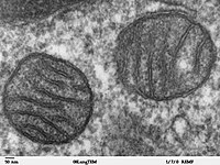
Photo from wikipedia
Alzheimer’s Disease (AD) is a common neurodegenerative disease that displays two hallmark protein accumulations in the brain – extracellular amyloid beta (Aβ) and intracellular hyperphosphorylated tau. Aβ has been shown… Click to show full abstract
Alzheimer’s Disease (AD) is a common neurodegenerative disease that displays two hallmark protein accumulations in the brain – extracellular amyloid beta (Aβ) and intracellular hyperphosphorylated tau. Aβ has been shown to directly penetrate the neuronal cell membrane and damage the membrane. This compromises membrane integrity, increases membrane permeability that alters membrane conductance and elevates intracellular calcium concentrations which contribute to reactive oxygen species production and mitochondrial dysfunction. This damage to the plasma membrane requires a quick and efficient repair mechanism to restore the barrier function of the membrane and avoid neuronal cell death. However, a defect in membrane repair has not been examined as a contributor to AD. Here, we aimed to assess membrane repair capacity in AD in vitro models as a potential contributor to neuronal death. To assess the effect of Aβ on membrane repair kinetics, we exposed primary mouse hippocampal neurons to 1μM recombinant human Aβ42 (rhAβ42) for 16 hours prior to experimentation. Additionally, we treated wildtype neuronal cells with healthy and AD patient with elevated levels of Aβ (PSEN1 M146L mutation) cortical neuron iPSC conditioned media. We also exposed primary mouse hippocampal neurons to cerebrospinal fluid (CSF) from non-AD and AD patients. Treated cells were challenged with an established laser damage assay where a two-photon laser is used to damage the cell membrane in the presence of FM4-64 dye. Dye fluorescence at the injury site is used as a proxy for membrane repair kinetics. Lastly, we investigated autoantibody production targeting proteins involved in membrane repair in non-AD and AD sera and CSF via custom ELISAs. Overall, we observed an induced membrane repair defect when wildtype neuronal cells are treated with rhAβ42, AD iPSC conditioned media, and AD CSF. We also observed a significant increase in autoantibody production in AD biofluids as compared to the non-AD controls. Autoantibody levels were inversely correlated with efficient membrane repair capacity and positively correlated with Aβ concentrations. These results illustrate Aβ’s role in impairing neuronal membrane repair capacity, and the potential contribution of autoantibodies targeting membrane repair proteins in AD. This work is supported by The Ohio State University Dean's Discovery grant and Dept. of Physiology & Cell Biology Nishikawara Student Research grant. This is the full abstract presented at the American Physiology Summit 2023 meeting and is only available in HTML format. There are no additional versions or additional content available for this abstract. Physiology was not involved in the peer review process.
Journal Title: Physiology
Year Published: 2023
Link to full text (if available)
Share on Social Media: Sign Up to like & get
recommendations!