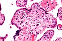
Photo from wikipedia
Introduction: During pregnancy, the maternal heart undergoes physiological cardiac growth, which reverses following parturition; however, the underlying processes that contribute are largely unknown. Changes in both systemic and cardiac metabolism… Click to show full abstract
Introduction: During pregnancy, the maternal heart undergoes physiological cardiac growth, which reverses following parturition; however, the underlying processes that contribute are largely unknown. Changes in both systemic and cardiac metabolism appear to be associated with cardiac growth, which could also contribute during pregnancy. We have shown that several cardiac metabolic transcripts and proteins and liver-derived metabolites are elevated during pregnancy and postpartum and are associated with cardiac growth. Therefore, the goal of this study was to examine the extent to which the cardio-hepatic signaling axis contributes to pregnancy-induced cardiac growth and reversal. Methods and Results: Timed pregnancy studies in 12-week-old FVB/NJ mice revealed that both heart weight (HW:TL; p < 0.0001) and liver weight (LIV:TL; p <0.0001) were highest in lactating 1-week post-birth (PB) mice versus mid-pregnant (8 d), late-pregnant (LP; 16 d), and non-pregnant (NP) mice. Interestingly, we also found that HW:TL ( p < 0.01) and LIV:TL ( p < 0.0001) were significantly reduced in non-lactating 1-week post-birth mice (PBNL) compared with PB mice. Hearts and livers both remained significantly larger, irrespective of lactation status, following parturition through 6 weeks post birth (PB) versus NP. Plasma proteomics revealed significant increases in prolactin at LP in addition to increases in liver-derived proteins including hepatocyte growth factors; however, these levels returned to NP levels by PB. Examination of the liver proteome from the same mice revealed significant increases in fatty acid and ketone body metabolism proteins. Cardiac and liver immunoblot analysis revealed significant reductions in Pdk4 between 1- and 3-week PB and PBNL groups that was reversed by 6-weeks postpartum and a significant increase in Pdk4 expression at 6 weeks postpartum, respectively. Interestingly, we see a sustained increase in cardiac Bdh1 expression that persisted at 6-weeks postpartum which could contribute to the sustained increases in HW:TL versus NP. Conclusion: Changes in both lactation status and metabolism are likely contributing to similar patterns of growth seen in the heart and liver during and following pregnancy. NIGMS COBRE Pilot project grant P30 GM127607; NIH R01 HL147844 (Hill/ Jones); Jewish Heritage Fund for Excellence Faculty Support; Jewish Heritage Fund for Excellence Research Enhancement Award; UofL School of Medicine Basic Science award; NIGMS IDeA state funded proteomics voucher. This is the full abstract presented at the American Physiology Summit 2023 meeting and is only available in HTML format. There are no additional versions or additional content available for this abstract. Physiology was not involved in the peer review process.
Journal Title: Physiology
Year Published: 2023
Link to full text (if available)
Share on Social Media: Sign Up to like & get
recommendations!