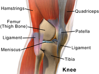
Photo from wikipedia
Introduction The aim of this study was to investigate entry points for anterior ankle arthroscopy that would minimize the risk of neurovascular injury. Methods Thirty-eight specimens from 21 Korean cadavers… Click to show full abstract
Introduction The aim of this study was to investigate entry points for anterior ankle arthroscopy that would minimize the risk of neurovascular injury. Methods Thirty-eight specimens from 21 Korean cadavers (age range from 43 to 92 years, mean age of 62.3 years) were used for the study. For the measurements, the most prominent points of the lateral malleolus (LM) and the medial malleolus (MM) were identified before dissection. A line connecting the LM and MM, known as the intermalleolar line, was used as a reference line. We measured 14 variables passed on the reference line. Results This study found that the nerves were located at 40.0%, 50.0%, and 82.0% of the reference line from the lateral malleolus. We also found that the arteries were located at 22.0%, 35.0%, and 60% of the reference line from the lateral malleolus. Discussion If all the variables are combined (nerves, arteries, and veins), then there is no safety zone for anterior portal placement. Therefore, we recommend that surgeons concentrate primarily on the arteries and nerves in the clinical setting.
Journal Title: BioMed Research International
Year Published: 2018
Link to full text (if available)
Share on Social Media: Sign Up to like & get
recommendations!