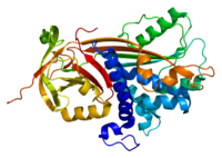
Photo from wikipedia
Ocular ischemia/hypoxia is a severe problem in ophthalmology that can cause vision impairment and blindness. However, little is known about the changes occurring in the existing fully formed choroidal blood… Click to show full abstract
Ocular ischemia/hypoxia is a severe problem in ophthalmology that can cause vision impairment and blindness. However, little is known about the changes occurring in the existing fully formed choroidal blood vessels. We developed a new whole organ culture model for ischemia/hypoxia in rat eyes and investigate the effects of pigment epithelium derived factor (PEDF) protein on the eye tissues. The concentration of oxygen within the vitreous was measured in the enucleated rat eyes and living rats. Then, ischemia was mimicked by incubating the freshly enucleated eyes in medium at 4°C for 14 h. Eyes were fixed immediately after enucleation or were intravitreally injected with PEDF protein or with vehicle before incubation. After incubation, light and electron microscopy (EM) as well as Tunel staining was performed. In the living rats, the intravitreal oxygen concentration was on average at 16.4% of the oxygen concentration in the air and did not change throughout the experiment whereas it was ca. 28% at the beginning of the experiment and gradually decreased over time in the enucleated eyes. EM analysis revealed that the shape of the choriocapillaris changed dramatically after 14 h incubation in the enucleated eyes. The endothelial cells made filopodia-like projections into the vessel lumen. They appeared identical to the labyrinth capillaries found in surgically extracted choroidal neovascular membranes from patients with wet age-related macular degeneration (AMD). These filopodia-like projections nearly closed the vessel lumen and showed open gaps between neighboring endothelial cells. PEDF significantly inhibited labyrinth capillary formation and kept the capillary lumen open. The number of TUNEL-positive ganglion cells and inner nuclear layer cells was significantly reduced in the PEDF-treated eyes compared to the vehicle-treated eyes. The structural changes in the chroidal vessels observed under ischemia/hypoxia conditions can mimic early changes in the process of pathological angiogenesis as observed in wet AMD patients. This new model can be used to investigate short-term drug effects on the choriocapillaris after ischemia/hypoxia and it highlighted the potential of PEDF as a promising candidate for treating wet AMD.
Journal Title: Oxidative Medicine and Cellular Longevity
Year Published: 2022
Link to full text (if available)
Share on Social Media: Sign Up to like & get
recommendations!