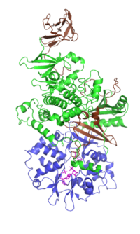
Photo from wikipedia
Purpose Oral squamous cell carcinoma (OSCC) is the sixth leading cause of cancer-related death worldwide and is characterized by metastasis and recurrence. We aimed to evaluate the expression of AKT1… Click to show full abstract
Purpose Oral squamous cell carcinoma (OSCC) is the sixth leading cause of cancer-related death worldwide and is characterized by metastasis and recurrence. We aimed to evaluate the expression of AKT1 and PLK1 in OSCC and identify their correlation with the clinical and histological features and prognosis of patients with OSCC. Methods Tissue samples were collected from 70 patients with OSCC and 50 patients with normal oral mucosa. The expression levels of AKT1 and PLK1 in OSCC tissues and normal oral mucosa were detected by immunohistochemistry. The chi-square test was used to identify correlations between the expression levels of AKT1 and PLK1 with patients' clinicopathologic characteristics. Survival analysis was assessed by the Kaplan–Meier method. Spearman's rank correlation test was used to determine the relationships between AKT1 and PLK1 expressions. The bioinformatics database GEPIA was used to verify the experimental results. Results The chi-square test and Fisher's exact test showed that the positive expression rate of AKT1 and PLK1 in OSCC tissue was significantly higher than that in the normal oral mucosa (P < 0.05). PLK1 expression levels were significantly correlated with tumor stage and size (P < 0.05). Kaplan–Meier analysis showed that the survival time of AKT1 and PLK1 with high expression was significantly shorter than that of patients with low expression (P < 0.05). Spearman's rank correlation test showed a strong correlation between AKT1 and PLK1 expression in OSCC tissue (R = 0.53; P < 0.05). GEPIA bioinformatics database analysis results show that the expression and overall survival of AKT1 and PLK1 analysis and the correlation analysis of AKT1 and PLK1 were consistent with experimental results. Conclusion AKT1 and PLK1 expressions are associated with the occurrence and progression of OSCC and may be used as diagnostic and prognostic indicators of OSCC. There may be a correlation between AKT1 and PLK1 in OSCC tissue.
Journal Title: Disease Markers
Year Published: 2022
Link to full text (if available)
Share on Social Media: Sign Up to like & get
recommendations!