Photo from wikipedia
Purpose: To examine whether ultra-widefield (UWF) retinal imaging can identify biomarkers for Alzheimer’s disease (AD) and its progression. Methods: Images were taken using a UWF scanning laser ophthalmoscope (Optos P200C… Click to show full abstract
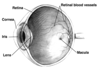
Purpose: To examine whether ultra-widefield (UWF) retinal imaging can identify biomarkers for Alzheimer’s disease (AD) and its progression. Methods: Images were taken using a UWF scanning laser ophthalmoscope (Optos P200C AF) to determine phenotypic variations in 59 patients with AD and 48 healthy controls at baseline (BL). All living participants were invited for a follow-up (FU) after 2 years and imaged again (if still able to participate). All participants had blood taken for genotyping at BL. Images were graded for the prevalence of age-related macular degeneration-like pathologies and retinal vascular parameters. Comparison between AD patients and controls was made using the Student t test and the χ2 test. Results: Analysis at BL revealed a significantly higher prevalence of a hard drusen phenotype in the periphery of AD patients (14/55; 25.4%) compared to controls (2/48; 4.2%) [χ2 = 9.9, df = 4, p = 0.04]. A markedly increased drusen number was observed at the 2-year FU in patients with AD compared to controls. There was a significant increase in venular width gradient at BL (zone C: 8.425 × 10⁻³ ± 2.865 × 10⁻³ vs. 6.375 × 10⁻³ ± 1.532 × 10⁻³, p = 0.008; entire image: 8.235 × 10⁻³ ± 2.839 × 10⁻³ vs. 6.050 × 10⁻³ ± 1.414 × 10⁻³, p = 0.004) and a significant decrease in arterial fractal dimension in AD at BL (entire image: 1.250 ± 0.086 vs. 1.304 ± 0.089, p = 0.049) with a trend for both at FU. Conclusions: UWF retinal imaging revealed a significant association between AD and peripheral hard drusen formation and changes to the vasculature beyond the posterior pole, at BL and after clinical progression over 2 years, suggesting that monitoring pathological changes in the peripheral retina might become a valuable tool in AD monitoring.
Journal Title: Ophthalmic Research
Year Published: 2018
Link to full text (if available)
Share on Social Media: Sign Up to like & get
recommendations!