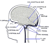
Photo from wikipedia
Abstract We report a rare case of recurrent isolated internal ophthalmoplegia attributed to oculomotor nerve (CN III) compression by the posterior cerebral artery (PCA). A 30-year-old female patient presented with… Click to show full abstract
Abstract We report a rare case of recurrent isolated internal ophthalmoplegia attributed to oculomotor nerve (CN III) compression by the posterior cerebral artery (PCA). A 30-year-old female patient presented with recurrent right-sided headaches, right periorbital pain, and slight anisocoria. Slit-lamp examination revealed normal anterior and posterior segments except for vermiform movements of the right pupil with a temporal hyporeactive flat area. Tonic pupils were ruled out with pilocarpine 0.1% testing. Suspecting an internal ophthalmoplegia, magnetic resonance imaging was ordered which demonstrated the right CN III indented by the PCA, fulfilling the criteria of a neurovascular conflict. The evaluation of unilateral mydriasis from internal ophthalmoplegia should prompt neuroimaging with exclusion of aneurysmal or compressive lesions. CN III palsy can rarely be caused by vascular anatomical variants because of the proximity of the posterior intracranial circulation and CN III. Newer, more precise imaging techniques will better help characterize neurovascular conflicts presenting as cranial nerve palsies.
Journal Title: Case Reports in Ophthalmology
Year Published: 2023
Link to full text (if available)
Share on Social Media: Sign Up to like & get
recommendations!