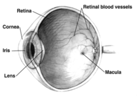
Photo from wikipedia
The structural and functional alterations of microvessels are detected because of physiological aging and in several cardiometabolic diseases, including hypertension, diabetes, and obesity. The small resistance arteries of these patients… Click to show full abstract
The structural and functional alterations of microvessels are detected because of physiological aging and in several cardiometabolic diseases, including hypertension, diabetes, and obesity. The small resistance arteries of these patients show an increase in the media or total wall thickness to internal lumen diameter ratio (MLR or WLR), often accompanied by endothelial dysfunction. For decades, micromyography has been considered as a gold standard method for evaluating microvascular structural alterations through the measurement of MLR or WLR of subcutaneous small vessels dissected from tissue biopsies. Micromyography is the most common and reliable method for assessing microcirculatory endothelial function ex vivo, while strain-gauge venous plethysmography is considered the reference technique for in vivo studies. Recently, several noninvasive methods have been proposed to extend the microvasculature evaluation to a broader range of patients and clinical settings. Scanning laser Doppler flowmetry and adaptive optics are increasingly used to estimate the WLR of retinal arterioles. Microvascular endothelial function may be evaluated in the retina by flicker light stimulus, in the finger by tonometric approaches, or in the cutaneous or sublingual tissues by laser Doppler flowmetry or intravital microscopy. The main limitation of these techniques is the lack of robust evidence on their prognostic value, which currently reduces their widespread use in daily clinical practice. Ongoing and future studies will overcome this issue, hopefully moving the noninvasive assessment of the microvascular function and structure from bench to bedside.
Journal Title: Hypertension
Year Published: 2022
Link to full text (if available)
Share on Social Media: Sign Up to like & get
recommendations!