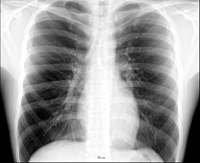
Photo from wikipedia
A 14-year-old previously healthy, fully vaccinated male presented with 3 weeks of intermittent left lower lateral chest wall pain and 3 days of fever and night sweats. The pain was… Click to show full abstract
A 14-year-old previously healthy, fully vaccinated male presented with 3 weeks of intermittent left lower lateral chest wall pain and 3 days of fever and night sweats. The pain was described as “pressure-like” that worsened when laying on the left side and painful to touch. He was seen at an urgent care while on a family trip approximately 1 week prior to admission where a chest x-ray reportedly showed a “lung infection.” He was prescribed a 5-day course of azithromycin. Over the next few days, he began to develop low-grade fevers with night sweats, prompting return to the hospital. The remaining review of systems was notable for 1 month of weight loss and negative for cough, congestion, shortness of breath, rash, fatigue, myalgia, or headache. He took no medications and had no known allergies. There was no family history of recurrent infection, autoimmune disease, or malignancy. He lived with his parents and younger brother in Southern California. He traveled to Colima, Mexico, 1 year prior to visit family, an area endemic for tuberculosis (TB) per Centers for Disease Control and Prevention (CDC) but had no known TB contacts. There were no pets in the home, no exposure to exotic animals or farm animals, and no recent sick contacts. On examination, he was an alert, thin-appearing male in no distress. His oropharynx was clear without erythema or exudates, and he had good dentition without evidence of caries or infection. There was no lymphadenopathy appreciated on palpation of the cervical neck region, axilla, or groin. He was stable on room air with symmetric chest rise, with decreased breath sound over the left lower lung field, and mild tenderness to touch over left lower rib cage. There was no chest wall deformity, swelling, or erythema. He had no appreciable organomegaly and no rash. His initial labs were notable for elevation in inflammatory markers (C-reactive protein 6.2 mg/dL, erythrocyte sedimentation rate 97 mm/h), with slight anemia to 11.3 g/dL, white blood cell of 10.9K/μL, and platelet count of 519K/μL. A complete metabolic panel, including liver function studies, was within normal limits. His initial chest x-ray demonstrated a patchy, nodular opacity in the left lung base. He underwent computed tomography of the chest and abdomen which showed a left lower lung consolidation (Figure 1) and a 6-cm mass-like structure (Figure 2) in the chest wall with periostitis and erosion of the 9th and 10th ribs, with direct invasion of the spleen. He was seen by the Infectious Diseases (ID) team and Oncology team. The initial infectious lab workup and a focused oncologic workup were negative. An interventional radiology (IR)-guided biopsy of the mass was inconclusive, and an open-incision biopsy was consistent with an infectious process. The patient underwent a video-assisted thoracic surgery and left lower lobe wedge resection, with filamentous bacteria and microabscesses seen on pathology, consistent with Actinomyces species.
Journal Title: Clinical Pediatrics
Year Published: 2022
Link to full text (if available)
Share on Social Media: Sign Up to like & get
recommendations!