Photo from wikipedia
Summary We postulate that blue telangiectasia and brownish pigmentation at ankle level, early markers of chronic venous insufficiency, can be quantified for longitudinal studies of chronic venous disease in Caucasian… Click to show full abstract
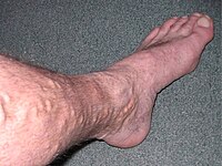
Summary We postulate that blue telangiectasia and brownish pigmentation at ankle level, early markers of chronic venous insufficiency, can be quantified for longitudinal studies of chronic venous disease in Caucasian people. Objectives and methods To describe a photographic technique specially developed for this purpose. The pictures were acquired using a dedicated photo stand to position the foot in a reproducible way, with a normalized lighting and acquisition protocol. The image analysis was performed with a tool developed using algorithms optimized to detect and quantify blue telangiectasia and brownish pigmentation and their relative surface in the region of interest. To test the short-term reproducibility of the measures. Results The quantification of the blue telangiectasia and of the brownish pigmentation using an automated digital photo analysis is feasible. The short-term reproducibility is good for blue telangiectasia quantification. It is a less accurate for the brownish pigmentation. Conclusion The blue telangiectasia of the corona phlebectatica and the ankle flare can be assessed using a clinimetric approach based on the automated digital photo analysis.
Journal Title: Phlebology
Year Published: 2018
Link to full text (if available)
Share on Social Media: Sign Up to like & get
recommendations!