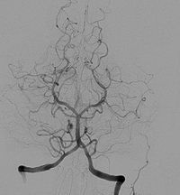
Photo from wikipedia
Background A non-invasive, reliable imaging modality that characterizes cavernous sinus dural arteriovenous fistula (CSDAVF) is beneficial for diagnosis and to assess resolution on follow-up. Purpose To assess the utility of… Click to show full abstract
Background A non-invasive, reliable imaging modality that characterizes cavernous sinus dural arteriovenous fistula (CSDAVF) is beneficial for diagnosis and to assess resolution on follow-up. Purpose To assess the utility of 3D time-of-flight (TOF) and silent magnetic resonance angiography (MRA) for evaluation of CSDAVF from an endovascular perspective. Material and Methods This prospective study included 37 patients with CSDAVF, who were subjected to digital subtraction angiography (DSA) and 3-T MR imaging with 3D TOF and silent MRA. The main arterial feeders, fistula site, and venous drainage pattern were evaluated, and the results were compared with DSA findings. The diagnostic confidence scores were also recorded using a 4-point Likert scale. Results Silent MRA correlated better for shunt site localization and angiographic classification (86% vs. 75% and 83% vs. 75%, respectively) compared to TOF MRA. The proportion of arterial feeders detected was marginally significant for silent MRA over TOF MRA sequences (92.8% vs. 89.5%; P=0.048), though for veins both were comparable. Sensitivity of silent MRA was higher for identification of cortical venous reflux (CVR) (90.9% vs. 81.8%) and deep venous drainage (82.4% vs. 64.7%), while specificity was >90% for both modalities. The overall diagnostic confidence score fared better for silent MRA for venous assessment (P < 0.001) as well as fistula point identification (P < 0.001), while no significant difference was evident with TOF MRA for arterial feeders (P=0.06). Conclusion Various angiographic components of CSDAVF could be identified and delineated by 3D TOF and silent MRA, though silent MRA was superior for overall diagnostic assessment.
Journal Title: Acta Radiologica
Year Published: 2022
Link to full text (if available)
Share on Social Media: Sign Up to like & get
recommendations!