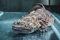
Photo from wikipedia
The authors describe pathological and microbiological features of mortalities in a captive breeding colony of Lord Howe Island stick insects (Dryococelus australis) over a period of 18 months. There were… Click to show full abstract
The authors describe pathological and microbiological features of mortalities in a captive breeding colony of Lord Howe Island stick insects (Dryococelus australis) over a period of 18 months. There were 2 peaks of mortality in this period. In the first, insects presented dead with minimal premonitory signs of illness. In the second, affected insects were ataxic with contracted limbs and inability to climb or right themselves. Gross lesions were uncommon but included pigmented plaques on the gut and cloacal prolapse. Histological lesions in both outbreaks indicated a cellular innate immune response including nodulation characterized by Gram-negative bacterial bacilli entrapped within nodules of pigmented hemocytes, and melanization characterized by melanin within hemocyte nodules and around bacteria. Hemolymph culture findings varied and often yielded a mixed growth. Pure growth of Serratia marcescens was cultured in 44% of animals in Outbreak 1, while pure growth of Pseudomonas aeruginosa was cultured in 30% of animals in Outbreak 2. Cases with S. marcescens-positive culture often showed inflammation at the foregut-midgut junction. The frequency of mixed bacterial culture results did not allow firm conclusions about causality to be made, and may indicate primary bacterial infection or increased susceptibility to hemolymph colonization with an opportunistic pathogen. These findings highlight the utility of histopathology combined with ancillary testing when investigating mortality in captive insect colonies.
Journal Title: Veterinary Pathology
Year Published: 2018
Link to full text (if available)
Share on Social Media: Sign Up to like & get
recommendations!