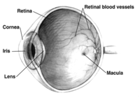
Photo from wikipedia
Objective This study aimed to investigate subclinical retinal microvascular changes with optical coherence tomography angiography (OCTA) in patients with systemic lupus erythematosus (SLE) and healthy controls (HCs), and to evaluate… Click to show full abstract
Objective This study aimed to investigate subclinical retinal microvascular changes with optical coherence tomography angiography (OCTA) in patients with systemic lupus erythematosus (SLE) and healthy controls (HCs), and to evaluate the relationship between OCTA findings and Systemic Lupus Erythematosus Disease Activity Index 2000 (SLEDAI-2K). Materials and Methods In this study, 47 eyes of SLE and 41 eyes of healthy control (HC) were evaluated. The SLE patients were divided into two subgroups: low disease activity (LDA) (SLEDAI≤5) and high disease activity (HDA) (SLEDAI>6). The results of OCTA were compared between SLE patients and HCs as well as the SLE subgroups. The relationship between OCTA results and SLEDAI-2K was evaluated. Results There were no differences in foveal avascular zone (FAZ) areas between the SLE patients and HCs. Central foveal thickness (CFT) was lower in SLE patients (p = .046). Superficial capillary plexus (SCP) vessel density (VD) in SLE patients was significantly lower only in the foveal area compared to that in HCs (p = .006). Deep capillary plexus (DCP) VD in SLE patients was significantly lower in all areas except the temporal parafoveal area compared to that in the HCs. There was no statistically significant difference between SLE groups with LDA and HDA in FAZ or any of the other areas, including SCP and DCP. When the correlation between OCTA findings and SLEDAI-2K was examined, both SCP and DCP VD were found to be negatively correlated. conclusions It was observed that DCP VDs were affected in SLE patients with LDA, and SCP VDs were also affected in addition to DCP with HDA. This suggests that DCP may be the first capillary plexus to be comprised in SLE. VDs were negatively correlated with disease activity. It was concluded that OCTA can be a useful tool in assessing subclinical retinal microvascular pathology and disease activity in patients with SLE.
Journal Title: Lupus
Year Published: 2022
Link to full text (if available)
Share on Social Media: Sign Up to like & get
recommendations!