Photo from wikipedia
BACKGROUND To evaluate the functional development and, retinal and optic disc morphology using OCT in patients with septo-optic dysplasia (SOD). METHODS This retrospective case series included patients diagnosed with SOD… Click to show full abstract
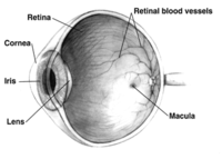
BACKGROUND To evaluate the functional development and, retinal and optic disc morphology using OCT in patients with septo-optic dysplasia (SOD). METHODS This retrospective case series included patients diagnosed with SOD between 2007 and 2020. Ophthalmologic assessment included visual acuity (VA) and funduscopy at the initial and last presentation. Retinal imaging included OCT of the macula analyzing the retinal morphology, central retinal thickness volume (CRT) and ganglion cell layer (GCL). Also, scans of the optic nerve head were taken to evaluate the retinal nerve fiber layer (RNFL) and global value. RESULTS 38 eyes of 19 children with a mean age 6.3 ± 5.3 years were included. 31.6% showed all 3 characteristics of SOD, whereof ONH, midline defects and endocrine dysfunctions were found in 94.7%, 89.5% and 47.4% respectively. The mean VA was 0.70 ± 0.66logMar in the right eye (RE) and 0.40 ± 0.55logMar in the left eye (LE) at the initial presentation. No change of vision (RE: 0.69 ± 0.71logMar; LE: 0.31 ± 0.57logMar) was found after a follow-up period of 6.3 ± 4.5years. Funduscopy showed an ONH in 79% (n = 30/38), tortuous retinal vessels in 36.8% (n = 14/38) and a double-ring sign in 15.8% (n = 6/38). Retinal imaging showed variable morphology. 6 eyes of 4 patients showed temporal retinal thinning with corresponding GCL attenuation. The optic nerve head appearance varied between no changes, sectoral and hemispherical reduction. CONCLUSIONS Patients suffering from SOD show diverse expression of retinal changes such as retinal, GCL and RNFL thinning in OCT. Furthermore, visual function remained stable during follow-up examinations, indicating no further alteration due to underlying pathology.
Journal Title: European journal of ophthalmology
Year Published: 2022
Link to full text (if available)
Share on Social Media: Sign Up to like & get
recommendations!