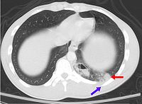
Photo from wikipedia
Severe lung damage is a major cause of death in blast victims, but the mechanisms of pulmonary blast injury are not well understood. Therefore, it is important to study the… Click to show full abstract
Severe lung damage is a major cause of death in blast victims, but the mechanisms of pulmonary blast injury are not well understood. Therefore, it is important to study the injury mechanism of pulmonary blast injury. A model of lung injury induced by blast exposure was established by using a simulation blast device. The effectiveness and reproducibility of the device were investigated. Eighty mice were randomly divided into eight groups: control group and 3 h, 6 h, 12 h, 24 h, 48 h, 7 days and 14 days post blast. The explosive device induced an explosion injury model of a single lung injury in mice. The success rate of the model was as high as 90%, and the degree of lung injury was basically the same under the same pressure. Under the same conditions, the thickness of the aluminum film can be from 0.8 mm to 1.6 mm, and the peak pressure could be from 95.85 ± 15.61 PSI to 423.32 ± 11.64 PSI. There is no statistical difference in intragroup comparison. A follow-up lung injury experiment using an aluminum film thickness of 1.4 mm showed a pressure of 337.46 ± 18.30 PSI induced a mortality rate of approximately 23.2%. Compared with the control group (372 ± 23 times/min, 85.9 ± 9.4 mmHg, 4.34 ± 0.09), blast exposed mice had decreased heart rate (283 ± 21 times/min) and blood pressure (73.6 ± 3.6 mmHg), and increased lung wet/dry weight ratio(2.67 ± 0.11), marked edematous lung tissue, ruptured blood vessels, infiltrating inflammatory cells, increased NF-κB (4.13 ± 0.01), TNF-α (4.13 ± 0.01), IL-1β (2.43 ± 0.01) and IL-6 (4.65 ± 0.01) mRNA and protein, decreased IL-10(0.18 ± 0.02) mRNA and protein (P < 0.05). The formation of ROS and the expression of MDA5 (4.46 ± 0.01) and IREα (3.43 ± 0.00) mRNA and protein were increased and the expression of SOD-1 (0.28 ± 0.02) mRNA and protein was decreased (P < 0.05). Increased expression of Bax (3.54 ± 0.00) and caspase 3 (4.18 ± 0.01) mRNA and protein inhibited the expression of Bcl-2 (0.39 ± 0.02) mRNA and protein. The changes of pulmonary edema, inflammatory cell infiltration, and cell damage factor expression increased gradually with time, and reached the peak at 12–24 h after the outbreak, and returned to normal at 7–14 days. Detonation injury can lead to edema of lung tissue, pulmonary hemorrhage, rupture of pulmonary vessels, induction of early inflammatory responses accompanied by increased oxidative stress in lung tissue cells and increased apoptosis in mice experiencing blast injury. The above results are consistent with those reported in other literatures. It is showed that the mouse lung blast injury model is successfully modeled, and the device can be used for the study of pulmonary blast injury. Impact statement The number of patients with explosive injury has increased year by year, but there is no better treatment. However, the research on detonation injury is difficult to carry out. One of the factors is the difficulty in making the model of blast injury. The laboratory successfully developed and produced a simulation device of explosive knocking through a large amount of literature data and preliminary experiments, and verified the preparation of the simulation device through various experimental techniques. The results showed that the device could simulate the shock wave-induced acute lung injury generated, which was similar to the actual knocking injury. The experimental process was controlled. Under the same condition, there was no statistical difference between the groups. It is possible to realize miniaturization and precision of an explosive knocking simulation device, which is a good experimental tool for further research on the mechanism of organ damage caused by detonation and the development of protective drugs.
Journal Title: Experimental Biology and Medicine
Year Published: 2018
Link to full text (if available)
Share on Social Media: Sign Up to like & get
recommendations!