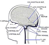
Photo from wikipedia
A 36-year-old female presented with severe left temporal headaches and a scalp mass with swelling and tenderness to palpation for 1 year. There was no history of trauma. A magnetic… Click to show full abstract
A 36-year-old female presented with severe left temporal headaches and a scalp mass with swelling and tenderness to palpation for 1 year. There was no history of trauma. A magnetic resonance angiography showed a focal 6-mm ovoid region of prominent flow-related enhancement along the course of the deep temporal artery (Figure 1). A cerebral angiogram showed an arteriovenous fistula supplied by the left middle deep temporal artery with venous drainage into the left superficial temporal vein that eventually drains into the left external jugular vein (Figure 2A and B). The lesion was completely embolized with n-Butyl cyanoacrylate (Figure 2C and D). At 8-month clinical follow-up, patient is completely asymptomatic.
Journal Title: Vascular and Endovascular Surgery
Year Published: 2019
Link to full text (if available)
Share on Social Media: Sign Up to like & get
recommendations!