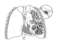
Photo from wikipedia
Since the outbreak of the COVID-19 pandemic, increasing evidence suggests that infected patients present a high incidence of thrombotic complications. We report a 67-year-old-woman admitted for severe acute respiratory syndrome… Click to show full abstract
Since the outbreak of the COVID-19 pandemic, increasing evidence suggests that infected patients present a high incidence of thrombotic complications. We report a 67-year-old-woman admitted for severe acute respiratory syndrome coronavirus 2 infection. Chest CT images showed bilateral ground glass opacities, bilateral pulmonary embolism, right ventricular clot in transit and 2 thoracic aortic mural thrombus. Therapy was initiated with subcutaneous low-molecular-weight heparin, and the patient was discharged at 20 days asymptomatic. Complete resolution of the aortic thrombus was observed in a 1-month surveillance CT angiogram. Our case illustrates vascular complications in a COVID-19 patient and its effective treatment with anticoagulation.
Journal Title: Vascular and Endovascular Surgery
Year Published: 2020
Link to full text (if available)
Share on Social Media: Sign Up to like & get
recommendations!