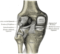
Photo from wikipedia
Background: Predictive factors influencing outcomes after surgical fixation of osteochondral fractures (OCFs) in the knee, particularly time between injury and surgery, have not been determined. Purpose: To report imaging and… Click to show full abstract
Background: Predictive factors influencing outcomes after surgical fixation of osteochondral fractures (OCFs) in the knee, particularly time between injury and surgery, have not been determined. Purpose: To report imaging and clinical outcomes after OCF fixation and to assess the association between clinical scores and patient characteristics, lesion morphology, and appearance on magnetic resonance imaging (MRI) scans. Study Design: Case series; Level of evidence, 4. Methods: We assessed the clinical and imaging outcomes of 19 patients after screw fixation for OCFs in the knee at a minimum follow-up of 1 year. Patient characteristics, lesion morphology, and time from trauma to surgery were reviewed for each patient. At final follow-up, patients completed a 100-point visual analog scale (VAS) for pain, Tegner activity scale, Knee injury and Osteoarthritis Outcome Score (KOOS), and patient satisfaction survey. Postoperative MRI scans were assessed using the MOCART (magnetic resonance observation of cartilage repair tissue), Osteochondral Allograft MRI Scoring System, and bone marrow edema (BME) size. Results: The mean patient age at surgery was 21.3 ± 11.4 years, and the median time from trauma to surgery was 10 days (range, 0-143 days). The refixed OCF fragment failed in 1 (5.3%) patient on the lateral condyle at 15 months postoperatively. The mean follow-up for the remaining 18 patients was 4.7 ± 3.2 years, and postoperative outcomes were as follows: VAS pain score, 9.5 ± 17.9; Tegner score, 4.8 ± 2.3; KOOS–Pain, 85.9 ± 17.6, KOOS-Symptoms, 76.4 ± 16.1; KOOS–Activities of Daily Living, 90.3 ± 19.0; KOOS–Sport, 74.4 ± 25.4; and KOOS–Quality of Life, 55.9 ± 24.7. Overall, 84.2% were satisfied or very satisfied with outcomes. Patient age was significantly associated with KOOS subscale scores and subchondral imaging parameters including BME and presence of subchondral cysts, which in turn were the only imaging variables linked to clinical outcomes (P < .05). Time from injury to surgery was not correlated with clinical or imaging outcomes. Conclusion: Fixation of OCFs yielded acceptable clinical and imaging outcomes at a mean 5-year follow-up with seemingly little influence of delayed surgical treatment. Postoperative subchondral changes were significantly associated with clinical outcomes and were linked to patient age at surgery.
Journal Title: Orthopaedic Journal of Sports Medicine
Year Published: 2022
Link to full text (if available)
Share on Social Media: Sign Up to like & get
recommendations!