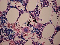
Photo from wikipedia
The mechanisms responsible for platelet generation have been debated for more than a century. It is generally assumed that the physiological release of platelets from megakaryocytes (MKs) occurs pri-marily by… Click to show full abstract
The mechanisms responsible for platelet generation have been debated for more than a century. It is generally assumed that the physiological release of platelets from megakaryocytes (MKs) occurs pri-marily by the release of platelet-sized swellings from the tips of proplatelets. 1,2 Proplatelets are long, branching membrane structures that extend from the MK surface into sinusoidal blood vessels in the bone marrow. They are also prominent in the lungs, and both MK and proplatelet fragmentation are thought to make a major contribution to in vivo platelet production. Much of the current understanding of proplatelet formation has been based on the characterization of cultured MKs, however it is notable that the structure and molecular mechanisms regulating in vitro proplatelet formation can differ signi fi cantly from what occurs in vivo. 3,4 A recent elegant study from Samir Taoudi ’ s laboratory 5 has provided comprehensive assessments of in vivo platelet biogenesis using whole-organ 3D and 4D quantitative imaging techniques during mouse embryogenesis, fetal development, and adult life. This study demonstrated that proplatelet formation in the bone marrow is uncommon (<5% of MKs), with most platelet-sized particles generated from distinct membrane structures, termed MK buds. Through a direct measurement of bud release at the whole-organ level, this study has challenged the long-held belief that proplatelets are predominant MK membrane structures generating platelets in vivo. 5 The MK budding theory remains controversial. Italiano et al have raised legitimate concerns as to whether buds contain the requisite structural features of mature platelets, including α -granules, an open
Journal Title: Blood Advances
Year Published: 2022
Link to full text (if available)
Share on Social Media: Sign Up to like & get
recommendations!