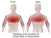
Photo from wikipedia
Introduction: In pediatric population pulmonary embolism(PE) is seen relatively rare compared to adults, but the incidence is increasing with the developed diagnostic methods. It has high morbidity and mortality when… Click to show full abstract
Introduction: In pediatric population pulmonary embolism(PE) is seen relatively rare compared to adults, but the incidence is increasing with the developed diagnostic methods. It has high morbidity and mortality when not correctly and timely diagnosed. Computerized Tomographic (CT) Angiography is the primary method used in PE. We aim to present the demographics, risk factors and therapies of 8 pulmonary emboli patients that we followed in our clinic in the last 1 year. Case: Pulmonary emboli was found in 8 cases; 2 were girls and the age range was 6-25. Of these 8 patients, 2 patients were following up with cystic fibrosis, 1 with duchenne muscular dystrophy, 1 with spina bifida who has mediastinal mass diagnosed as teratoma, 1 had fallot tetralogy, 1 was diagnosed as systemic lupus erythematosis who was obese. Our last patient had an 11-hour bus journey before complaining of chest pain in his medical history. In other 2 patients, no underlying disease was detected. However, these two patients were smokers. 7 of our patients had complains of chest pain, 1 patient presented with hemoptysis, followed by chest pain. The diagnosis was confirmed by CT angiography. Enoxaparin sodium was used as a treatment with d-dimer level above normal in all cases. Conclusion: In childhood age group, pulmonary emboli should be considered as a differential diagnosis of acute chest pain and diagnostic examinations should be planned.
Journal Title: European Respiratory Journal
Year Published: 2018
Link to full text (if available)
Share on Social Media: Sign Up to like & get
recommendations!