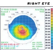
Photo from wikipedia
Background A larger optical zone for photorefractive keratectomy may improve optical quality and stability. However, there is need for limiting ablation diameter in that a larger ablation diameter requires greater… Click to show full abstract
Background A larger optical zone for photorefractive keratectomy may improve optical quality and stability. However, there is need for limiting ablation diameter in that a larger ablation diameter requires greater ablation depth, and minimizing ablation depth may reduce adverse effects on postoperative wound healing, haze and keratoectasia. In this study, we compared the changes in clinical outcomes and the degree of regression between a 6.0 mm optical zone and 6.5 mm optical zone following PRK. Methods The records of 95 eyes that had undergone PRK with a 6.0 OZ ( n = 40) and a 6.5 OZ ( n = 55) were retrospectively reviewed. We compared data including the spherical equivalent of manifest refraction (SE of MR), simulated K (Sim K), thinnest corneal thickness, change in thinnest corneal thickness (the initial value divided by corrected diopter [ΔTCT/CD]), Q value, corneal higher order aberrations (HOAs) and spherical aberration (SA) pre-operation, at 3 and 6 months postoperative and at the last follow-up visit (Mean; 20.71 ± 10.52, 17.47 ± 6.57 months in the 6.0 and 6.5 OZ group, respectively). Results There were no significant differences in the SE of MR, Sim K and UDVA between the 6.0 OZ group and the 6.5 OZ group over 1 year of follow-up after PRK, and the 6.0 OZ group required less ΔTCT/CD than the 6.5 OZ group. The 6.5 OZ group showed better results in terms of post-operative HOAs of RMS, SA and Q value. When comparing that pattern of change in Sim K, there was no significant difference between the 6.0 OZ group and the 6.5 OZ group. Conclusions The clinical refractive outcomes and regression after PRK using Mel 90 excimer laser with a 6.0 OZ were comparable to those with a 6.5 OZ.
Journal Title: BMC Ophthalmology
Year Published: 2020
Link to full text (if available)
Share on Social Media: Sign Up to like & get
recommendations!